Rating: 3.9/5
ACTEON X-MIND Optima 3D Review
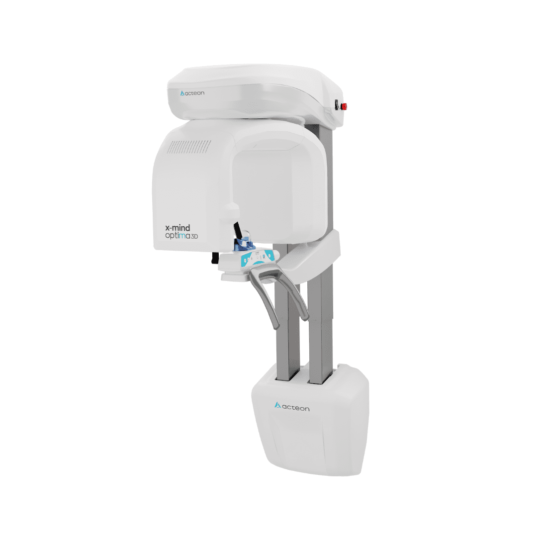
The ACTEON X-MIND Optima 3D is a panoramic and CBCT unit that mounts on a wall and weighs 69 kg. ACTEON unveiled it at the French Dental Association congress in Paris in November...
Reviewed by Agnes Markovic
Pros
- Wall-mounted unit weighs 69 kg and fits a 120 × 115 cm room
- Fields of view run from 5×5 cm up to an optional 17×12 cm
- IGZO detector with 16-bit depth produces clean, high-contrast volumes
- 90 kV ceiling improves penetration in heavily restored mouths
- 70 µm endo mode resolves fine canal anatomy on small volumes
- Every 3D program offers a low-dose option
- AIS and RealGUIDE PRO add AI nerve tracing and segmentation at no extra cost
- Open gantry accommodates standing, seated, and wheelchair patients
Cons
- Platform launched in late 2024, so long-term reliability data is limited
- Finest voxel of 70 µm trails units that advertise 50 µm
- DICOM networking and RealGUIDE DESIGN cost extra
- Ceph build loses the compact advantage at 126 kg
- No published effective dose figures for each program
- Acquisition PC needs a dedicated NVIDIA GPU and gigabit networking
- Service quality depends on local dealer coverage
The ACTEON X-MIND Optima 3D is a panoramic and CBCT unit that mounts on a wall and weighs 69 kg. ACTEON unveiled it at the French Dental Association congress in Paris in November 2024. This review looks at image quality, software, clinical range, and the costs that sit outside the quote.
TL;DR
The wall-mounted Optima weighs 69 kg and fits a room as small as 120 × 115 cm.
An IGZO detector and a 90 kV ceiling drive detailed scans, with a 70 µm endo mode for small volumes.
Fields of view span 5×5 cm to an optional 17×12 cm, so one unit covers single teeth and full maxillofacial volumes.
ACTEON bundles AIS and RealGUIDE PRO for AI nerve tracing, while guide design and DICOM networking cost extra.
What Is The ACTEON X-MIND Optima 3D
The X-MIND Optima 3D is ACTEON's newest 2D/3D extraoral imaging unit. It is built by de Götzen, the group's Italian imaging company, and sold through ACTEON's distributor network. It combines panoramic radiography with CBCT scanning in one gantry, and an optional cephalometric arm turns it into a full orthodontic imaging unit.
ACTEON positions the Optima as its most compact CBCT to date. The wall-mounted version needs no base plate, which explains the "zero-footprint" label on the company's X-MIND Optima 3D product page. A floor-standing version followed for rooms where the wall cannot carry the load, and ACTEON's US hub listed it for the first quarter of 2025.
The target buyer is a general or implant-focused practice adding its first 3D unit, or replacing an older panoramic machine without rebuilding the room. Specialists with heavy endodontic or full-skull caseloads should read the image quality and comparison sections closely before deciding.
Key Specifications At A Glance
The figures below come from ACTEON's North American technical datasheet, dated September 2024. Configuration codes and availability differ between countries, so confirm both with your local distributor.
Specification | Wall-mounted Pan/CBCT | Floor-standing Pan/CBCT | Pan/Ceph/CBCT |
|---|---|---|---|
Tube voltage | 60–90 kV | 60–90 kV | 60–90 kV |
Anode current | 2–12.5 mA | 2–12.5 mA | 2–12.5 mA |
Focal spot | 0.5 mm | 0.5 mm | 0.5 mm |
3D detector | IGZO flat panel, 110 µm pixel | IGZO flat panel, 110 µm pixel | IGZO flat panel, 110 µm pixel |
Ceph detector | Not applicable | Not applicable | CMOS with CsI scintillator, 99 µm pixel |
Gray levels | 16-bit (65,536) | 16-bit (65,536) | 16-bit 3D, 14-bit ceph |
Voxel size | 70 µm endo, 160 µm standard | 70 µm endo, 160 µm standard | 70 µm endo, 160 µm standard |
3D fields of view | 5×5 to 12×10 cm, 17×12 cm optional | 5×5 to 12×10 cm, 17×12 cm optional | 5×5 to 12×10 cm, 17×12 cm optional |
Exposure time | Pan up to 14 s, 3D from 5.3 s | Pan up to 14 s, 3D from 5.3 s | Ceph from 4.3 s |
Footprint | 1107 × 1063 mm | 1204 × 1063 mm | 1204 × 1859 mm |
Weight | 69 kg | 89 kg | 126 kg |
Software | AIS 5.4, RealGUIDE 5.4 PRO | AIS 5.4, RealGUIDE 5.4 PRO | AIS 5.4, RealGUIDE 5.4 PRO |
The 3D program list covers full dentition at 85×93 mm, a single jaw at 85×50 mm, and single-sextant volumes at 50×50 mm. It adds TMJ at 85×90 mm, maxillary sinuses at 85×93 mm, full arch and airways at 120×100 mm, and the extended arch at 170×120 mm. Every 3D program has a low-dose variant.
The 2D menu includes adult and child panoramic, low-dose panoramic, half panoramic, frontal dentition, orthoradial panoramic, bitewings, lateral TMJ in open and closed positions, and a PA sinus view. The ceph arm adds lateral and AP skull projections in several sizes, plus a 24×18 cm carpus view for skeletal age.
Design, Footprint, And Installation
Room size decides more CBCT purchases than image quality does, and this is where the Optima stands apart. The wall-mounted unit has a 1107 × 1063 mm footprint and needs a minimum room of 120 × 115 cm, with 160 × 150 cm recommended.
Many floor-standing 2D/3D units in this class weigh well over 100 kg. A 69 kg wall unit usually avoids a floor reinforcement survey, although the wall still needs a load assessment and proper anchoring. Practitioners in leased buildings should check the lease before drilling into a structural wall.
Three Configurations For Different Rooms
ACTEON sells the Optima in three builds, each available with a 12×10 cm base field of view or the extended 17×12 cm view.
Wall-mounted Pan/CBCT at 69 kg, the most compact build and the default for general and implant practices
Floor-standing Pan/CBCT at 89 kg, for glass partitions, thin stud walls, or leases that forbid wall mounting
Floor-standing Pan/Ceph/CBCT at 126 kg, with a 1650 mm ceph arm and a 1204 × 1859 mm footprint
The open gantry lets patients walk in and stand or sit without stepping onto a platform. Wheelchair users can usually stay in their own chair, which removes the transfer that slows down many CBCT appointments for older patients.
Planning The Room, Network, And Workstation
Installation planning starts with radiation protection. Most jurisdictions require a shielding plan and a post-install survey before the first patient scan, and operator credentialing rules vary by state, as our X-ray certification guide explains.
Layout matters as much as shielding. The operator needs a clear sightline to the patient during exposure, and the acquisition PC should sit within view of that position. Our guide to dental operatory design covers sightlines and traffic flow for imaging rooms.
The datasheet sets firm minimums for the acquisition workstation:
Intel Core i7 at 3 GHz, or a 13th-generation Core i5 or newer
At least 8 GB of RAM and a 1 TB hard disk
NVIDIA graphics with at least 4 GB of memory
Windows 10 64-bit and a free PCIe x16 slot for the supplied dual-port network card
ACTEON specifies a 1 Gb network connection for CBCT data. Review stations can run Windows or macOS, which helps practices where the front desk and the operatories use different platforms.
Image Quality And Diagnostic Performance
The Optima pairs an IGZO flat-panel detector with a 60–90 kV generator and a dedicated 70 µm endo mode. These choices put its imaging closer to mid-range CBCT than its size suggests, with caveats worth understanding before a demo.
What The IGZO Detector Changes
IGZO stands for indium gallium zinc oxide, a thin-film transistor material that is replacing amorphous silicon in newer X-ray panels. IGZO transistors move charge faster and leak less current, which lowers electronic noise and speeds up readout.
Independent physics work supports the general claim. A University of Milan characterization of an IGZO flat panel tested under CBCT exposure conditions reported a linear response and low image lag, both relevant to a rotating acquisition. That study used a radiotherapy panel, so it describes the technology class and not Optima's specific detector.
On the Optima, the panel has a 169.3 × 169.3 mm active area, 110 µm pixels, and 16-bit depth. The 65,536 gray levels help when one volume needs windowing for dense cortical bone and for the thin buccal plate over an anterior root.
Why A 90 kV Ceiling Matters
Higher tube voltage produces a harder beam that penetrates dense bone and metal more evenly. ACTEON's earlier X-MIND prime 3D tops out at 86 kV, so the Optima's 90 kV ceiling is a meaningful step for heavily restored mouths.
Beam hardening around crowns, posts, and implants produces the dark streaks that hide crestal bone and fracture lines. A higher kV setting reduces those streaks without removing them. The datasheet does not list a dedicated metal artifact reduction algorithm, so ask the distributor to scan a restored case during the demo.
Voxel Size And What It Means For Endodontics
The Optima reconstructs at 70 µm in endo mode and 160 µm in standard mode. The 70 µm setting is competitive for a compact unit and suits canal morphology, calcified canals, resorption, and periapical lesions on small volumes.
Voxel size is a reconstruction setting, and true spatial resolution also depends on detector noise, focal spot size, and patient stillness. A 2025 systematic review on vertical root fracture detection found that smaller voxels improved detection in teeth with metal posts in laboratory studies, while intracanal materials reduced accuracy overall. Clinicians chasing a suspected fracture should pair the endo mode with careful clinical testing, as described in our article on cracked tooth syndrome.
The AAE and AAOMR joint position statement on CBCT in endodontics recommends limited field of view scans for complex cases and the smallest field that answers the clinical question. The Optima's 5×5 cm endo volume fits that guidance, and it pairs well with the clinical signs listed in our guide to failed root canal symptoms.
Fields Of View From Single Tooth To Full Skull
The field of view range runs from 5×5 cm to an optional 17×12 cm (diameter × height), one of the widest spans in the compact category.
Field of view (Ø × H) | Typical clinical use |
|---|---|
5×5 cm | Endodontics, single implant site, localized pathology |
8.5×5 cm | Single jaw implant sites, impacted third molars |
8.5×9 cm | Full dentition, maxillary sinus, one TMJ |
12×10 cm | Full arches, airway screening, multiple implants |
17×12 cm (optional) | Both TMJs, orthodontic records, full-arch guided surgery |
The 17×12 cm view is a license activation, so a practice can buy the 12×10 cm build and unlock the larger volume later without new hardware. Collimating down to 5×5 cm keeps the dose confined to the region under investigation.
Dose Management
Every 3D program on the Optima has a low-dose option, and the 2D menu includes a low-dose panoramic. Child presets adjust exposure automatically, and 3D exposure times start at 5.3 seconds.
The FDA guidance on dental cone-beam CT notes that CBCT generally delivers more radiation than conventional dental X-rays and asks clinicians to justify each scan. The ADA's 2024 radiography recommendations went further on shielding, advising practices to stop routine use of thyroid collars and lead abdominal aprons while ordering images in moderation.
ACTEON's public materials do not list effective dose in microsieverts for each program. Practices that need dosimetry figures for local regulators should request them in writing before purchase.
Software And AI Workflow
Software is where the Optima earns much of its value. ACTEON ships the unit with Acteon Imaging Suite (AIS) 5.4 and RealGUIDE 5.4 PRO, and its US Optima hub describes the workstation, monitor, and software as included with no recurring fees.
Acteon Imaging Suite As The Hub
Acteon Imaging Suite stores panoramic, CBCT, intraoral sensor, and camera images in one patient record. Practices already using ACTEON sensors or phosphor plates gain a single database in place of several vendor applications.
AIS handles multiplanar views, curved panoramic reconstruction from the volume, measurements, annotations, and exports. ACTEON's newer pages reference AIS 5.5, so confirm which version the installer will load.
RealGUIDE AI Tools
RealGUIDE is a cloud-connected planning suite that ZimVie also sells for guided surgery and restorative design. Inside AIS, it adds AI tools aimed at the slowest parts of implant planning.
One-click detection of the inferior alveolar canal
Assisted bone segmentation for printed models and guide design
Automatic tooth segmentation with virtual extraction
AI-assisted alignment of intraoral STL scans to the CBCT volume
ZimVie's RealGUIDE 5.4 brochure claims nerve detection drops from about 5 minutes to under 1 minute, and full-arch bone segmentation from about 1.5 hours to roughly 2 minutes. These are manufacturer figures. The nerve trace still needs slice-by-slice review, particularly where the canal runs close to the lingual cortex or loses its corticated border.
Our overview of AI in dentistry explains where automated segmentation helps and where it fails. The report on FDA-cleared dental AI for CBCT lists which tools carry regulatory clearance, and our guide to dental imaging AI compares how vendors apply it.
What Is Included And What Costs Extra
The North American datasheet separates the standard bundle from paid options.
Item | Status |
|---|---|
AIS 5.4 acquisition and viewing | Included |
RealGUIDE 5.4 PRO planning | Included |
RealGUIDE 5.4 DESIGN for guide and restorative design | Optional, annual renewal |
Extended 17×12 cm field of view | Optional activation, bundled in some promotions |
DICOM package (Print, Worklist, Store, Query/Retrieve, DSR) | Optional |
Practices that refer scans to radiologists or hospitals will want the DICOM package for PACS integration. Check separately that DICOM exports open cleanly in any third-party planning software already in use.
Clinical Applications By Specialty
Fields of view from 5×5 to 17×12 cm let the Optima cover nearly every indication a general or specialist practice images in-house. The sections below map the programs to daily clinical work.
Implant Planning And Guided Surgery
The AAOMR position statement on implant imaging recommends cross-sectional imaging for all implant sites and names CBCT as the method of choice. Implant planning is Optima's core use case.
The 8.5×5 cm single-jaw volume covers most single and multiple implant sites, and the 12×10 cm volume suits full-arch cases. Bone width, cortical thickness, the mental foramen, and the sinus floor all become measurable, which matters when a case may need a sinus lift or ridge augmentation.
RealGUIDE merges the volume with an intraoral scanner file so the implant position follows the planned crown. A practice with in-house 3D printing can then print the surgical guide chairside. Our guides to guided implantology and implant treatment planning walk through each stage, and the immediate versus early implant loading article covers how bone quality on the scan shapes the loading protocol.
Endodontics
The 5×5 cm endo volume at 70 µm targets missed canals, calcifications, resorption, and periapical lesions hidden on periapical films.
A dedicated endodontic practice imaging single teeth all day may prefer a unit with a smaller voxel. For a general practice doing its own endodontic triage, the endo mode adds certainty that periapicals cannot provide, particularly on maxillary molars with a suspected MB2 canal.
Oral Surgery And Third Molars
CBCT clarifies how an impacted wisdom tooth relates to the mandibular canal, which shapes both the surgical approach and the consent discussion.
The 8.5×5 cm volume handles a single third molar well. Larger cysts, impacted canines, and supernumerary teeth may need the 12×10 cm view.
Orthodontics And Airway
The ceph build adds cephalometric X-ray projections and a carpus view for skeletal age assessment.
The 12×10 cm airway program and the 17×12 cm view support 3D records for impacted canines, root resorption, and alveolar bone limits before aligner or bracket movement. Airway volumes from a dental scan are screening information only, and suspected sleep-disordered breathing needs a medical referral.
TMJ And Sinus Assessment
Dedicated TMJ (85×90 mm) and sinus (85×93 mm) programs sit alongside 2D lateral TMJ and PA sinus views.
The 17×12 cm volume captures both condyles in one scan, which helps compare joint morphology in patients with suspected TMJ disorder. CBCT does not show the articular disc, so MRI remains the reference for internal derangement.
Clinical Walkthrough: A Representative Implant And Endo Day
The scenario below is an illustrative composite built from Optima's published programs and a typical implant workflow. It shows how the unit fits a clinical day and does not report a controlled trial or a specific patient.
A 58-year-old patient arrives for a consult on a missing lower left first molar, extracted eight months earlier. The assistant selects the 8.5×5 cm single-jaw program in standard mode. The patient walks into the open gantry, bites on the positioning block, and holds still for an exposure that starts at 5.3 seconds. Patients who remember older panoramic units often remark on how quickly the arm finishes.
The volume opens in AIS within minutes. RealGUIDE traces the inferior alveolar canal, and the clinician checks the trace on cross-sections where the canal flattens near the mental foramen. In this scenario, the ridge measures about 7 mm at the crest, enough for a regular-diameter implant without grafting. The team merges an intraoral scan, places a virtual crown, and positions the implant beneath it with a 2 mm margin above the canal.
Case presentation is where the 3D view earns its place. The patient sees the nerve and the planned implant on screen, with the bone wrapped around both, and that picture tends to settle worries about numbness faster than a verbal explanation. Our article on patient communication in dentistry covers how to structure that conversation. The findings then go into a written dental treatment plan with phases, fees, and the surgical guide as a line item.
Later the same day, a patient with lingering pain after root canal therapy on an upper first molar gets a 5×5 cm scan in endo mode. The axial slices show an untreated MB2 canal and a small radiolucency at the mesiobuccal apex. Neither finding was visible on the periapical film, and the retreatment plan now has a clear target.
In a day like this, the Optima contributes speed and the confidence of seeing anatomy in three planes. Its limits show up in heavily restored mouths, where streaks from adjacent crowns can still obscure fine detail at 70 µm.
Workflow Tips For Consistent Scans
Consistent CBCT images depend more on positioning and protocol than on hardware. The habits below apply to the Optima and to most compact units.
Select the smallest field of view that answers the clinical question
Stabilize the head carefully, since motion during rotation causes most repeat scans
Remove earrings, partial dentures, and piercings from the scanned area
Use cotton rolls to separate the arches when occlusal surfaces matter
Reserve endo mode for fine detail and use standard mode for implant planning
Run the acquisition PC on a dedicated wired gigabit connection
Training shapes results as well. ACTEON publishes a 2D/3D positioning guide on its US hub and supports onboarding through its distributors. Its US materials state that third-party IT and dealer onsite fees are not ACTEON's responsibility, so confirm who handles installation and networking before signing.
How It Compares With Other CBCT Units
The Optima competes with compact 2D/3D units from Carestream, Vatech, and Planmeca, as well as ACTEON's own X-MIND prime 3D. Published headline specifications differ in ways that matter for specific practices.
Unit | Max field of view | Finest voxel | Mount and weight | Notable feature |
|---|---|---|---|---|
ACTEON X-MIND Optima 3D | 17×12 cm (optional) | 70 µm | Wall 69 kg or floor 89 kg | IGZO panel, bundled AI planning |
ACTEON X-MIND prime 3D | 12×10 cm | 87.5 µm | Floor, about 67 kg | Predecessor, 86 kV maximum |
Carestream CS 8200 3D Advance | 16×10 cm (top edition) | 75 µm | Floor | Upgradeable FOV editions |
Vatech Green X | 18×15 cm | 50 µm | Floor | Compressed-sensing reconstruction |
Planmeca ProMax 3D Classic | 8×8 cm single scan | 75 µm | Floor, 113 kg | Romexis software ecosystem |
The Optima leads on the wall mount, the 17×12 cm option at its price level, and bundled AI planning. Units advertising 50–75 µm voxels may edge ahead for an endodontic practice, and larger brands often have more service engineers in a given region.
For deeper looks at alternatives, see our Carestream CS 9600 review, our Planmeca Viso G7 review for full-face imaging, our analysis of the DEXIS OP 3D LX, and our Planmeca Romexis review for the software side.
Price And Cost Of Ownership
ACTEON does not publish list prices for the Optima. Quotes depend on configuration, field of view license, software options, installation, and region.
According to ACTEON's US hub, the bundle includes the acquisition workstation, monitor, AIS, and RealGUIDE PRO with no recurring software fees. Extra costs usually come from shielding work, the DICOM package, RealGUIDE DESIGN renewals for in-house guide design, and extended warranty. Some regional promotions have bundled the 17×12 cm view, so ask whether one applies.
ACTEON also offers an ROI calculator on its US hub to model payback from in-house scans and implant cases. Our dental clinic financing guide compares leasing and loan options for imaging equipment.
Bottom Line
The ACTEON X-MIND Optima 3D puts a 17×12 cm field of view and AI-assisted implant planning into a 69 kg unit that hangs on a wall. Many practices have postponed CBCT because the only free space is an old panoramic bay, and this unit changes that calculation. The room can usually stay as it is, and the software needed to plan an implant arrives with the scanner.
Image quality sits closer to mid-range CBCT than the unit's size suggests. The IGZO panel and 90 kV ceiling handle full-arch implant work and most endodontic questions well. Fine detail around crowns and posts is where the limits show, and a demo scan of a heavily restored case is the quickest way to judge whether those limits matter for a given patient base.
The fit is narrower for specialists. An endodontic practice that images single teeth all day may get more from a unit advertising a 50 µm voxel. Orthodontists choosing the ceph build give up most of the compact advantage, since it weighs 126 kg and needs 1859 mm of floor depth.
Before signing, buyers should get per-program dose figures and local service response times in writing, and price DICOM and RealGUIDE DESIGN in the first quote. Those items shape the cost of ownership more than the base price does. The 17×12 cm view unlocks later as a license, so a practice unsure about orthodontic or TMJ volumes can start with the 12×10 cm build.
Verdict
<p>For a general practice adding its first CBCT, or an implant practice replacing an aging small-volume unit, the ACTEON X-MIND Optima 3D is one of the most sensible choices in the compact category. It addresses the constraint most practices face, which is room size. A 69 kg wall mount that fits a former panoramic bay removes the construction work that delays many CBCT purchases.</p><p>Clinically, the pairing of an IGZO detector with a 90 kV ceiling covers nearly every indication a general practice images in-house, from a suspected MB2 canal to a full-arch guided case. The bundled RealGUIDE tools deserve more attention than they usually get. Automated nerve tracing and STL merging shorten implant planning to minutes, provided the clinician still checks every slice.</p><p>The reservations are modest. The platform is young, so practices should ask for local references and service response times in writing. Endodontists imaging single teeth all day may want a finer voxel, and orthodontists should plan for the larger ceph footprint. DICOM and DESIGN licenses belong in the first quote.</p><p>Practices that need 3D imaging in a small room should put the Optima on their demo list.</p>
Frequently Asked Questions
What is the ACTEON X-MIND Optima 3D?
It is a combined panoramic and CBCT unit from ACTEON, manufactured by its Italian subsidiary de Götzen. It launched in November 2024 and comes as a wall-mounted unit, a floor-standing unit, or a floor-standing unit with a cephalometric arm.
How much space does the X-MIND Optima 3D need?
The wall-mounted version needs a minimum room of 120 × 115 cm, with 160 × 150 cm recommended. The ceph build needs much more depth because of its 1204 × 1859 mm footprint, so a site survey is always advised.
What fields of view does it offer?
Standard builds cover 5×5 cm up to 12×10 cm. An optional extended view reaches 17×12 cm (diameter × height) and can be activated after installation.
What is the voxel size of the X-MIND Optima 3D?
Endo mode reconstructs at 70 µm and standard mode at 160 µm. Endo mode suits small volumes for canal anatomy and fractures, while standard mode suits implant planning and larger volumes.
Is the X-MIND Optima 3D good for implant planning?
Implant planning is its strongest use. It ships with AIS and RealGUIDE 5.4 PRO, which add AI nerve detection, bone and tooth segmentation, STL merging, and implant positioning. Surgical guide design requires the optional RealGUIDE DESIGN module.
Can orthodontists use it?
Orthodontists can use the ceph build, which adds lateral and AP skull projections plus a carpus view. The 17×12 cm view and the airway program support 3D orthodontic records.
How long does a scan take?
3D exposures start at 5.3 seconds, a standard panoramic takes up to 14 seconds, and ceph exposures start at 4.3 seconds.
How does it differ from the X-MIND prime 3D?
The Optima raises the kV ceiling from 86 to 90 kV and moves to an IGZO flat panel. It also reduces the finest voxel from 87.5 to 70 µm and extends the maximum field of view from 12×10 to 17×12 cm, in a lighter design that can mount on a wall.
Does it work with third-party software?
The unit runs on Acteon Imaging Suite. An optional DICOM package adds Print, Worklist, Store, Query/Retrieve, and DSR for PACS and hospital networks. Confirm export compatibility with your preferred planning software before purchase.
How much does the X-MIND Optima 3D cost?
ACTEON prices the Optima by quote, and totals vary with configuration, software options, and region. In the US, the workstation, monitor, AIS, and RealGUIDE PRO come bundled, while installation, shielding, DICOM, and DESIGN renewals add to the total.