Rating: 4.5/5
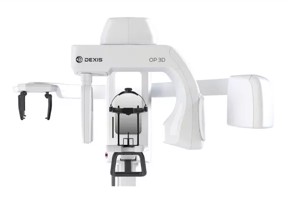
DEXIS Orthopantomograph™ OP 3D™ LX: A Comprehensive Review for Modern Dental Practices
Understanding the DEXIS OP 3D LX System in Today's Dental LandscapeIn today's competitive dental landscape, diagnostic imaging equipment can make or break a practice's ability to...
Reviewed by Rachel Thompson
Pros
- Comprehensive Multi-Modality Platform: Combines panoramic, cephalometric, and CBCT imaging in one unit, eliminating the need for multiple separate systems and maximizing space efficiency while simplifying staff training and equipment maintenance.
- Large Field-of-View Capability: The LX model offers extensive imaging volume up to 17 x 13 cm, ideal for full-arch implant planning, comprehensive orthodontic evaluation, maxillofacial assessment, and complex surgical cases requiring detailed anatomical visualization.
- Exceptional Image Quality: Delivers consistently sharp, detailed images across all modalities with excellent contrast, minimal artifacts, and high spatial resolution sufficient for demanding diagnostic applications.
- Advanced Dose Management: Multiple FOV options ranging from focused small-volume scans to full maxillofacial imaging, pulsed exposure modes, copper filtration, and automatic exposure control support radiation dose reduction while maintaining diagnostic image quality.
- User-Friendly Interface: Intuitive touchscreen operation with clear positioning guidance, real-time feedback on patient alignment, and straightforward protocol selection simplifies workflow and reduces training time for staff members.
- Robust Software Ecosystem: Seamless integration with DEXIS imaging software provides comprehensive tools for image enhancement, measurement, implant planning, cephalometric analysis, and annotation without requiring third-party applications.
- Excellent Interoperability: DICOM compatibility ensures smooth integration with major practice management systems, treatment planning software, and CAD/CAM platforms, supporting comprehensive digital workflows.
- Patient-Friendly Design: Open architecture reduces claustrophobia, relatively short scan times minimize patient discomfort, and comfortable positioning accessories accommodate patients ranging from children to large adults.
- True Multi-Modality Imaging: Provides authentic panoramic and cephalometric projections rather than synthetic reconstructions, maintaining familiar image appearance and diagnostic characteristics clinicians expect.
- Established Service Network: Backed by DEXIS support infrastructure with comprehensive service availability, regular software updates, and established parts supply chain for long-term reliability.
- Enhanced Practice Capabilities: Enables in-house imaging that would otherwise require referrals to imaging centers, improving patient convenience, practice profitability, and case acceptance while expanding service offerings.
- Versatile Clinical Applications: Supports diverse specialties from implant dentistry and endodontics to orthodontics, oral surgery, and TMJ assessment with appropriate imaging protocols for each application.
- Workflow Efficiency: Single platform handles multiple imaging needs, reducing patient appointments, eliminating equipment-switching delays, and simplifying staff training compared to managing multiple separate systems.
- Professional Consultation Enhancement: High-quality 3D reconstructions, cross-sectional views, and annotated images displayed on chairside monitors facilitate patient education and treatment acceptance.
- Scalable Resolution Options: Multiple resolution settings allow practices to prioritize maximum detail for complex cases or faster acquisition with lower dose for routine screening, optimizing the balance for each clinical situation.
- Quality Assurance Tools: Built-in QA features facilitate routine testing and documentation, supporting regulatory compliance and ensuring consistent image quality over the system's operational lifetime.
Cons
- Substantial Capital Investment: Purchase price typically ranging from $150,000-$200,000 represents a significant financial commitment that may challenge smaller practices, startups, or practices with limited capital budgets.
- Significant Space Requirements: Despite being compact for a multi-function unit, requires dedicated space approximately 2.0 x 2.4 meters plus additional clearance for patient access, operator positioning, and cephalometric arm extension.
- Potential Overkill for Specialized Practices: Single-specialty practices focused exclusively on one modality may not utilize all capabilities, making dedicated single-purpose systems potentially more cost-effective with lower initial investment.
- Installation Complexity: May require facility modifications including radiation shielding installation, electrical upgrades, dedicated circuit protection, and professional installation coordination adding to total project cost.
- Learning Curve for Advanced Features: While basic operation proves user-friendly, maximizing the system's advanced capabilities including CBCT interpretation and treatment planning requires comprehensive training and ongoing education.
- Ongoing Maintenance Costs: Long-term ownership includes annual preventive maintenance visits, service contract costs, software update fees, and potential repair expenses that impact total cost of ownership beyond purchase price.
- Software Licensing Dependencies: Optimal functionality requires compatible DEXIS software with potential additional licensing costs for advanced modules like implant planning or cephalometric analysis.
- Technology Evolution Risk: Rapid advances in imaging technology mean premium systems risk relative obsolescence as newer models with enhanced capabilities, improved dose efficiency, or AI-enhanced features emerge.
- Utilization Requirements: Significant investment requires adequate imaging volume across all modalities to achieve positive return on investment, potentially challenging for lower-volume practices.
- Regulatory Compliance Burden: Operating imaging equipment requires ongoing regulatory compliance including equipment registration, operator credentialing, quality assurance documentation, and radiation safety program maintenance.
- Limited Soft Tissue Visualization: Like all CBCT systems, primarily visualizes hard tissue anatomy with limited soft tissue contrast, requiring complementary imaging modalities for comprehensive soft tissue assessment.
- Dependence on Local Service: System performance and uptime depend significantly on local service availability and technician expertise, which may vary by geographic location.
- Electrical Infrastructure Requirements: Requires dedicated electrical circuits with appropriate voltage and amperage, potentially necessitating electrical panel upgrades in older facilities.
- Staff Training Investment: Achieving competency across all modalities requires time investment for initial training and continuing education, temporarily reducing productivity during the learning phase.
- Workflow Disruption During Installation: Installation process may require temporary closure of the designated room, impacting practice operations during the installation and setup period.
Understanding the DEXIS OP 3D LX System in Today's Dental Landscape
In today's competitive dental landscape, diagnostic imaging equipment can make or break a practice's ability to deliver exceptional patient care while maintaining operational efficiency and profitability. The DEXIS Orthopantomograph OP 3D LX emerges as a compelling solution for practices seeking to consolidate multiple imaging modalities into one sophisticated platform. But does this all-in-one approach truly deliver on its promise, or does it sacrifice specialized performance for versatility?
The dental imaging market has evolved dramatically over the past two decades. Gone are the days when a simple panoramic X-ray unit sufficed for most diagnostic needs. Modern dentistry demands detailed three-dimensional visualization for implant planning, comprehensive assessment of impacted teeth, precise endodontic diagnosis, and sophisticated orthodontic treatment planning. Patients increasingly expect their dental providers to offer advanced diagnostic capabilities in-house rather than referring them to separate imaging centers.
This convergence of clinical necessity and patient expectation has created a market opportunity for manufacturers to develop integrated imaging solutions. DEXIS, leveraging decades of expertise in dental imaging, has positioned the OP 3D LX as a answer to these evolving demands. The question for dental professionals becomes not whether to invest in advanced imaging, but which platform best serves their specific practice needs, patient demographics, and growth objectives.
The Evolution of DEXIS Imaging Technology
DEXIS has established itself as a prominent name in dental imaging, with a history spanning multiple decades and technological generations. The company's journey from intraoral sensors to comprehensive extraoral imaging systems reflects the broader evolution of digital dentistry. Understanding this heritage provides context for evaluating the OP 3D LX's position in the current marketplace.
The Orthopantomograph line has long been recognized for reliability and image quality in panoramic radiography. As CBCT technology matured and became more accessible to general dental practices, DEXIS strategically expanded the Orthopantomograph platform to incorporate three-dimensional imaging capabilities. This evolutionary approach, rather than creating entirely new product lines, allowed DEXIS to leverage existing expertise while meeting emerging clinical demands.
The OP 3D designation indicates the integration of cone beam computed tomography into the traditional panoramic platform. The addition of "LX" specifies this as the large field-of-view variant, distinguishing it from more compact versions designed for practices with more limited space or imaging requirements. This tiered product approach allows practices to select the configuration that best aligns with their clinical needs and physical constraints.
What Sets the OP 3D LX Apart from Competing Systems
The DEXIS OP 3D LX distinguishes itself through its multi-functional design philosophy. Unlike single-purpose imaging systems that require practices to invest in separate units for panoramic, cephalometric, and CBCT imaging, this system integrates all three modalities seamlessly. This consolidation represents not just a space-saving measure, but a fundamental rethinking of workflow efficiency in modern dental practices.
Competing systems in the market generally fall into three categories: dedicated panoramic units, dedicated CBCT systems, and integrated multi-function platforms. Dedicated units typically excel in their specific modality but require multiple equipment purchases and consume more operatory space. Some CBCT systems offer panoramic reconstruction from volumetric data, but these synthetic panoramic images often lack the sharpness and familiar appearance of true panoramic projections.
The OP 3D LX's approach provides authentic imaging in each modality rather than compromising on synthetic reconstructions. When a clinician selects panoramic mode, the system performs a true panoramic acquisition with the focal trough and projection geometry specific to that examination. This authenticity extends to cephalometric imaging, where dedicated lateral and PA projections provide the standardized views orthodontists expect for cephalometric analysis.
The "LX" designation indicates this is the large field-of-view variant, capable of capturing volumetric data across a substantial imaging area measuring up to 17 x 13 cm. This capability proves particularly valuable for practices handling complex cases involving full-arch implant planning, comprehensive orthodontic assessments requiring visualization of the entire maxillofacial complex, or surgical extractions requiring detailed anatomical visualization of adjacent structures.
Technical Capabilities and Imaging Performance
At the heart of any imaging system's value proposition lies its technical performance—the fundamental ability to capture diagnostic-quality images that reveal anatomical details with clarity and precision. The DEXIS OP 3D LX distinguishes itself through sophisticated engineering that balances multiple competing priorities: maximizing image quality while minimizing radiation exposure, providing versatile field-of-view options while maintaining consistent resolution, and delivering rapid acquisition times without compromising diagnostic detail. Understanding these technical capabilities requires examining each imaging modality individually, as the system's true strength emerges not from excelling in a single dimension but from maintaining exceptional performance across its entire range of functions. From the volumetric precision of its CBCT acquisitions to the familiar reliability of its panoramic imaging and the standardized geometry of its cephalometric projections, the OP 3D LX demonstrates that multi-modality design need not compromise specialized performance.
CBCT Imaging Excellence and Volumetric Acquisition
The cone beam computed tomography functionality of the OP 3D LX delivers high-resolution 3D imaging with multiple field-of-view options ranging from focused small-volume scans to full maxillofacial imaging. Practices can select from various scanning volumes depending on the clinical indication, optimizing the balance between image detail, scan time, and radiation exposure. This flexibility proves essential for adhering to ALARA principles while ensuring diagnostic adequacy.
The sensor technology employs a flat-panel detector that captures projection data during the rotational scan. This detector architecture provides excellent spatial resolution with minimal noise, contributing to the system's ability to visualize fine anatomical details such as the lamina dura, trabecular patterns, and the periodontal ligament space. The large format of the flat-panel detector enables the extended field-of-view capability that defines the LX model.
Image quality remains consistently sharp across different scanning protocols, with sophisticated reconstruction algorithms minimizing artifacts and enhancing diagnostic detail. The system employs advanced scatter correction techniques that reduce the beam-hardening artifacts commonly seen in CBCT imaging, particularly in the presence of high-density materials like amalgam restorations or implants. These artifact reduction capabilities prove crucial for accurate diagnosis and treatment planning.
The volumetric data acquisition typically completes within 14 to 18 seconds depending on the selected protocol, translating to relatively brief exposure times for patients. This swift acquisition minimizes the potential for motion artifacts while reducing patient anxiety associated with remaining still during the scan. For pediatric patients or those with difficulty maintaining static positions, these shorter scan times significantly improve the likelihood of diagnostic-quality acquisitions.
The system offers multiple resolution settings, allowing practices to prioritize either maximum detail for complex cases or faster acquisition with lower dose for routine screening examinations. High-resolution protocols provide voxel sizes as small as 0.075 mm, delivering the fine detail necessary for endodontic planning or evaluation of subtle pathology. Standard resolution protocols with voxel sizes around 0.15-0.20 mm suffice for most implant planning and general diagnostic applications while requiring less reconstruction time and storage space.
Panoramic Radiography for Daily Diagnostic Needs
Beyond 3D imaging, the OP 3D LX excels in traditional panoramic radiography, which remains the workhorse examination for screening, initial assessment, and routine monitoring. The panoramic programs include standard full-arch imaging along with specialized programs for pediatric patients, temporomandibular joint assessment, sinus evaluation, and focused views of specific jaw segments.
These programmed options streamline workflow by automatically adjusting exposure parameters, rotation timing, and focal trough dimensions based on the selected examination type. The system's patient positioning aids include laser alignment guides and anatomical landmarks displayed on the acquisition workstation, helping technicians achieve consistent, reproducible positioning that minimizes the need for retakes.
The panoramic image quality demonstrates excellent sharpness and contrast, providing clear visualization of dental structures, bone levels, and pathology. The dynamic exposure control adjusts radiation output during the scan based on tissue density, optimizing image quality while minimizing patient dose. This technology proves particularly valuable when imaging patients with asymmetric anatomy or varying tissue densities across the scan field.
One often-overlooked advantage of true panoramic acquisition versus reconstructed panoramic views from CBCT data lies in the familiar image appearance and consistent focal trough geometry. Clinicians trained on conventional panoramic radiography can interpret these images using established pattern recognition without adjusting to the different appearance of synthetic panoramic reconstructions. This familiarity reduces interpretation errors and maintains diagnostic confidence.
The panoramic modality also provides exceptional efficiency for screening examinations where 3D imaging isn't clinically indicated. A routine recall examination for a healthy patient requires only the brief exposure and quick acquisition of a panoramic view, preserving the 3D capability for cases where its diagnostic value justifies the additional radiation exposure and cost.
Cephalometric Capabilities for Orthodontic Excellence
For orthodontic practices or general practitioners offering orthodontic services, the integrated cephalometric functionality proves invaluable. The system captures lateral and posteroanterior cephalometric projections essential for orthodontic diagnosis and treatment planning. The cephalometric arm positions precisely, ensuring reproducible patient positioning for progress monitoring throughout treatment.
The lateral cephalometric program provides the standardized lateral skull radiograph orthodontists rely upon for cephalometric analysis. The system maintains consistent magnification factors and projection geometry, allowing for accurate linear and angular measurements. The image receptor size accommodates the full range of patient sizes from pediatric cases to large adults, ensuring complete anatomical coverage without compromising on detail.
Digital cephalometric imaging eliminates the variables associated with traditional film processing while enabling immediate analysis with compatible software platforms. This immediate access to diagnostic data accelerates treatment planning and enhances patient consultation experiences. Parents can view their child's cephalometric radiograph on a monitor during the consultation, with the orthodontist highlighting key findings and explaining the recommended treatment approach in real-time.
The posteroanterior cephalometric capability, while used less frequently than lateral projections, provides essential information for assessing facial asymmetry, transverse discrepancies, and certain types of dentofacial deformities. Having this capability integrated into the same platform that provides lateral cephalometric and CBCT imaging proves particularly valuable for complex orthodontic cases requiring comprehensive assessment.
Advanced orthodontic practices increasingly combine traditional 2D cephalometric analysis with 3D CBCT evaluation for comprehensive diagnosis. The OP 3D LX enables practices to perform both examinations during a single patient visit, streamlining the diagnostic process and reducing the number of appointments required before treatment initiation. This efficiency enhances patient satisfaction while optimizing practice productivity.
Workflow Integration and Software Ecosystem
The OP 3D LX integrates seamlessly with the DEXIS imaging software suite, providing an intuitive interface for image acquisition, processing, and analysis. The software offers robust tools for image enhancement, measurement, and annotation, supporting thorough diagnostic evaluation without requiring third-party applications. The measurement tools include standardized cephalometric analysis templates, implant planning modules, and endodontic measurement capabilities.
The acquisition workstation features a dedicated computer system running the DEXIS software, ensuring optimal performance and stability. The interface design emphasizes clarity and efficiency, with large, easily identifiable icons for different examination types and clear visual feedback during patient positioning. The touchscreen functionality allows operators to navigate through options and initiate scans without requiring a separate keyboard and mouse, reducing the physical footprint of the acquisition station.
For practices using comprehensive practice management systems, the OP 3D LX typically interfaces smoothly with major dental software platforms through standard DICOM protocols. This interoperability ensures captured images flow directly into patient records, maintaining organized documentation and supporting efficient clinical workflows. The DICOM compatibility extends to both sending images to the practice management system and retrieving patient demographic information to populate the acquisition fields automatically.
The image processing capabilities include adjustable brightness, contrast, and sharpness filters that allow clinicians to optimize the image appearance for their diagnostic preferences. The software also provides advanced tools such as distance and angle measurements, density measurements for bone quality assessment, and annotation features for marking anatomical structures or pathology. These tools enable thorough documentation and support clear communication with patients and referring doctors.
The CBCT viewer provides multi-planar reconstruction with synchronized scrolling through axial, coronal, and sagittal planes. The ability to navigate through the volumetric dataset in multiple orientations simultaneously proves invaluable for understanding three-dimensional anatomical relationships. The viewer also generates oblique slices along the dental arch, providing cross-sectional views perpendicular to the arch that simplify implant site assessment and endodontic evaluation.
The system's acquisition station features a user-friendly touchscreen interface that simplifies patient positioning and protocol selection. Even staff members new to 3D imaging can achieve consistent results after minimal training, thanks to the intuitive design and clear visual guidance provided during setup. The software provides real-time feedback on patient positioning, alerting operators if the patient's head is tilted or rotated beyond acceptable parameters before initiating the scan.
Patient Experience Considerations and Comfort Features
Modern patients increasingly value practices that invest in advanced diagnostic technology. The OP 3D LX's open design reduces the claustrophobic feeling some patients experience with older, more enclosed imaging systems. The unit's architecture provides clear sightlines for patients during positioning and acquisition, allowing them to see the surrounding room and maintain visual contact with the operator. This openness particularly benefits pediatric patients and adults with anxiety issues.
The chin rest and temple supports provide stable, comfortable positioning during acquisition. These patient contact points feature smooth, easily sanitized surfaces that facilitate infection control between patients. The adjustable height accommodation allows the system to position comfortably for patients ranging from small children to tall adults, ensuring proper alignment regardless of patient stature.
The relatively short scan times minimize patient discomfort and reduce the likelihood of motion artifacts compromising image quality. Patients appreciate spending less time in the unit, particularly when multiple imaging modalities are needed during a single visit. The ability to complete panoramic, cephalometric, and CBCT acquisitions without repositioning the patient between different machines significantly enhances the patient experience.
Radiation exposure remains a valid concern for health-conscious patients, particularly parents considering imaging for their children. The OP 3D LX addresses this through selectable field-of-view options and optimized exposure protocols. By imaging only the region of clinical interest and utilizing dose-reduction technologies, the system delivers diagnostic images while respecting ALARA (As Low As Reasonably Achievable) principles.
The system's dose-reduction technologies include pulsed exposure modes that deliver radiation only during optimal projection angles, copper filtration that removes low-energy photons that contribute to patient dose without improving image quality, and sophisticated automatic exposure control that adjusts radiation output based on patient size and tissue density. These combined technologies can reduce radiation exposure by 30-50% compared to older-generation CBCT systems while maintaining diagnostic image quality.
For practices that emphasize patient education as part of their care philosophy, the OP 3D LX supports enhanced consultation experiences. The ability to display three-dimensional reconstructions, cross-sectional views, and annotated images on chairside monitors helps patients understand their diagnosis and appreciate the rationale for recommended treatment. This visual communication often proves more effective than verbal explanations alone, particularly for complex procedures like implant placement or surgical extractions.
Space and Installation Requirements
The footprint of the OP 3D LX, while compact for a multi-function unit, still requires dedicated space within the practice. The unit itself occupies approximately 2.0 x 2.4 meters of floor space, though practices must account for additional clearance around the unit for patient access, operator positioning, and the extended reach of the cephalometric arm during lateral skull acquisitions.
Practices considering this system should evaluate their available space carefully, accounting not just for the unit itself but for the clearance needed for the rotating arm during cephalometric acquisitions. The rotating C-arm that holds the X-ray source and detector requires unobstructed space during its orbital path around the patient. Ceiling height requirements typically range from 2.4 to 2.7 meters depending on specific installation configurations.
The physical placement within the practice deserves careful consideration from both workflow and regulatory perspectives. Ideally, the unit should be positioned in a dedicated room that allows for controlled access during acquisitions. The room should provide sufficient space for an operator console positioned where the technician can monitor the patient visually during scans while maintaining appropriate distance from the primary beam.
Installation involves both the imaging unit and a separate acquisition workstation. The unit requires appropriate electrical connections, typically 230V single-phase power with dedicated circuit protection. The electrical requirements are generally modest compared to some imaging equipment, but practices should verify their existing electrical infrastructure can accommodate the system without requiring panel upgrades.
The unit may necessitate lead-lined walls or other radiation shielding depending on adjacent spaces and local regulations. A qualified radiation physicist or health physicist should perform a shielding analysis considering the types and volumes of examinations the practice anticipates performing, the construction of existing walls and floors, and the occupancy of adjacent spaces. This analysis determines whether existing construction provides adequate shielding or whether supplemental lead lining, lead-impregnated drywall, or other shielding solutions are necessary.
Working with experienced installation professionals ensures the system integrates properly into the practice environment while meeting all regulatory requirements. DEXIS typically partners with authorized installation specialists who understand the specific requirements of the OP 3D LX and can coordinate the various aspects of installation including electrical work, shielding modifications, network connectivity, and equipment positioning.
Clinical Applications Across Specialties
Technical specifications and image quality metrics ultimately matter only insofar as they translate into meaningful clinical value across the diverse range of cases that modern dental practices encounter daily. The true measure of the OP 3D LX's worth lies in its ability to enhance diagnostic confidence, improve treatment planning precision, and support superior patient outcomes across multiple dental disciplines. From the exacting demands of implant site assessment to the three-dimensional complexity of endodontic anatomy, from the comprehensive evaluation required for orthodontic diagnosis to the anatomical clarity essential for surgical planning, each specialty presents unique imaging requirements and diagnostic challenges. The versatility of a multi-modality platform proves its value when a single system can seamlessly transition from providing a routine panoramic screening for a hygiene patient to delivering detailed volumetric data for complex implant reconstruction, all within the same clinical day. The following sections explore how the OP 3D LX's imaging capabilities address the specific needs of major dental specialties, examining both the strengths that make it valuable and the limitations practitioners should understand when applying this technology to clinical practice.
Implant Dentistry and Surgical Planning
For implant cases, the OP 3D LX provides the detailed anatomical information essential for precise treatment planning. Clinicians can evaluate bone volume, assess bone quality through density measurements, identify vital structures like the inferior alveolar nerve and maxillary sinus, and plan implant positioning with confidence. The cross-sectional views perpendicular to the dental arch provide the classic "slice" images implant dentists rely upon for determining available bone height and width.
The ability to capture the full arch in a single scan streamlines full-mouth rehabilitation planning. Practices can visualize the entire maxilla or mandible volumetrically, allowing for comprehensive assessment of bone anatomy, identification of anatomic variations, and planning of multiple implant positions simultaneously. This comprehensive view proves particularly valuable when planning complex cases such as All-on-4 or other full-arch implant-supported prostheses.
The implant planning software modules that integrate with the DEXIS platform enable virtual implant placement within the CBCT dataset. Clinicians can position virtual implants, adjust their angulation, assess proximity to adjacent structures, and evaluate the amount of bone engagement. This virtual planning reduces surgical uncertainty and allows for preoperative identification of potential complications such as inadequate bone volume or proximity to anatomical structures.
For practices that utilize guided surgery protocols, the CBCT data from the OP 3D LX can be exported to surgical guide design software. The combination of CBCT data and intraoral scans or conventional impressions enables the fabrication of surgical guides that translate the virtual plan into precise surgical execution. This digital workflow represents the cutting edge of implant dentistry and requires the type of high-quality volumetric data the OP 3D LX provides.
The system's capability to perform both initial diagnostic CBCT scans and post-operative verification scans with consistent geometry and resolution supports comprehensive documentation of surgical outcomes. Practices can compare pre-operative and post-operative datasets to verify implant positioning, assess osseointegration, and document treatment results for quality assurance and medico-legal purposes.
Endodontics and Complex Root Canal Anatomy
Endodontists benefit from the system's ability to reveal complex root canal anatomy, including extra canals, calcifications, and periapical pathology that may be difficult to visualize on conventional two-dimensional radiographs. The three-dimensional perspective eliminates the superimposition that can obscure additional canals or unusual anatomic configurations on periapical radiographs.
The focused field-of-view options available on the OP 3D LX allow for high-resolution imaging of individual teeth or small groups of teeth. These small-volume scans deliver voxel sizes that approach the resolution of periapical radiographs while providing three-dimensional information. For endodontic applications, this combination of resolution and volumetric data proves ideal for identifying calcified canals, assessing root fractures, and evaluating the extent of periapical lesions.
The 3D perspective proves particularly valuable for retreatment cases where understanding why the initial treatment failed is essential for planning the revision approach. Clinicians can identify missed canals, assess the adequacy of previous obturation, visualize perforations or separated instruments, and evaluate the three-dimensional extent of periapical pathology. This comprehensive assessment supports more predictable retreatment outcomes.
The cross-sectional views provided by the CBCT dataset allow for precise measurement of canal dimensions, assessment of canal curvature in three dimensions, and identification of anatomic features such as C-shaped canals or taurodont roots. This detailed anatomic information enables endodontists to select appropriate instruments, plan their cleaning and shaping procedures, and anticipate technical challenges before beginning treatment.
For general dentists who perform endodontic therapy, the OP 3D LX provides a valuable tool for case selection and referral decisions. The ability to visualize the complete root canal anatomy before beginning treatment helps identify cases that exceed the practitioner's comfort level or technical capabilities, supporting appropriate referral to endodontic specialists while retaining straightforward cases in the general practice.
Orthodontics and Comprehensive Dentofacial Assessment
The combined cephalometric and CBCT capabilities support comprehensive orthodontic diagnosis that considers both dental and skeletal relationships. Clinicians can assess airway dimensions, evaluate impacted teeth, visualize root angulations, and plan treatment with a complete understanding of the three-dimensional relationships between dental and skeletal structures.
Airway assessment has become increasingly important in orthodontic treatment planning as the profession recognizes the relationship between craniofacial development, orthodontic treatment, and airway function. The OP 3D LX enables visualization and quantification of pharyngeal airway dimensions, identification of airway constrictions, and documentation of airway changes during orthodontic treatment. This capability proves particularly valuable for cases involving orthognathic surgery or rapid palatal expansion where airway implications require careful consideration.
The three-dimensional visualization of impacted teeth reveals their precise position relative to adjacent teeth, their relationship to vital structures like the maxillary sinus or inferior alveolar nerve, and their angulation relative to the dental arch. This information guides decisions about whether to extract impacted teeth or attempt surgical exposure and orthodontic alignment. The 3D data also facilitates communication with oral surgeons when planning combined surgical-orthodontic approaches to impacted teeth.
For practices that offer accelerated orthodontics or surgically-facilitated orthodontics, the CBCT capability enables assessment of alveolar bone thickness and morphology. Clinicians can evaluate whether adequate bone volume exists for proposed tooth movements, identify areas where bone grafting might be necessary, and plan corticotomy patterns for procedures like periodontally-accelerated osteogenic orthodontics.
The ability to perform virtual treatment planning using the CBCT dataset, particularly when combined with digital models from intraoral scanning, represents the future of orthodontic diagnosis and treatment planning. Practices can simulate tooth movements in three dimensions, evaluate the effect of proposed tooth positions on facial esthetics, and generate customized appliances based on the virtual treatment plan. The OP 3D LX provides the foundational diagnostic data that enables these advanced digital orthodontic workflows.
Oral Surgery, Extractions, and Pathology Assessment
Surgical cases involving impacted teeth, pathology, or trauma benefit enormously from the detailed visualization the OP 3D LX provides. Surgeons can plan surgical approaches, anticipate complications, and communicate treatment plans more effectively with referring doctors and patients. The three-dimensional perspective reveals the spatial relationships that determine surgical difficulty and guide technique selection.
For impacted third molars, the CBCT data reveals the tooth's relationship to the inferior alveolar canal, the configuration of the tooth's root anatomy, the density of surrounding bone, and the presence of any associated pathology such as dentigerous cysts. This comprehensive assessment allows surgeons to provide more accurate informed consent discussions with patients regarding the risks of nerve injury, estimate surgical difficulty, and plan the most appropriate surgical technique.
The assessment of jaw pathology benefits significantly from three-dimensional imaging. Cysts, tumors, and other lesions can be visualized in their entirety, their relationship to adjacent structures can be evaluated, and their effect on surrounding bone can be assessed. The ability to measure lesion dimensions accurately in three dimensions supports treatment planning and provides baseline data for monitoring lesion growth or regression during observation periods.
Trauma cases often involve complex fractures of the facial skeleton that are difficult to assess completely on two-dimensional radiographs. The CBCT capability of the OP 3D LX enables comprehensive evaluation of fracture patterns, displacement of fracture segments, and involvement of vital structures. This detailed assessment guides treatment planning and facilitates communication with oral and maxillofacial surgery colleagues or medical trauma teams.
For general dentists who provide surgical extractions or evaluate jaw pathology, the OP 3D LX enhances diagnostic confidence and supports appropriate case selection. The detailed visualization helps identify cases that require referral to specialists while providing the information necessary to manage straightforward surgical cases competently. This capability expands the scope of services general practices can provide while maintaining appropriate standards of care.
TMJ Assessment and Dysfunction Evaluation
The temporomandibular joint imaging capabilities of the OP 3D LX support evaluation of TMJ disorders, a common condition affecting a significant percentage of the population. The system provides dedicated TMJ programs that capture both joints simultaneously, enabling comparison between the right and left sides. This bilateral visualization often reveals asymmetries in joint morphology or degenerative changes that contribute to understanding the patient's symptoms.
The CBCT imaging reveals bony changes in the condyle and temporal fossa that may not be apparent on conventional TMJ radiographs or clinical examination alone. Clinicians can identify flattening of the condylar head, osteophyte formation, erosive changes, or asymmetric condylar morphology. These findings inform diagnosis and help differentiate between different categories of TMJ disorders such as osteoarthritis, degenerative joint disease, or developmental abnormalities.
The ability to assess joint space width and condylar position within the fossa provides information about disc displacement, though soft tissue structures like the articular disc itself are not directly visualized without contrast-enhanced imaging or MRI. Despite this limitation, the bony changes visible on CBCT often correlate with the severity and duration of disc displacement, providing clinically useful information for treatment planning.
For practices that provide TMJ treatment through occlusal appliances, orthodontics, or other therapeutic modalities, the OP 3D LX enables comprehensive documentation of joint status before treatment and monitoring of joint morphology during and after treatment. This documentation supports evaluation of treatment outcomes and provides important medico-legal documentation of the patient's condition.
Return on Investment Considerations and Financial Analysis
The financial commitment required for the OP 3D LX represents a significant practice investment, typically ranging from $150,000 to $200,000 depending on configuration, installation requirements, and negotiated terms. This capital outlay demands careful financial analysis to ensure the investment aligns with practice economics and delivers appropriate returns.
However, the multi-modality capability potentially eliminates the need for multiple separate imaging systems, consolidating capital expenditure. A practice equipped with separate panoramic, cephalometric, and CBCT units might have $250,000 or more invested across these systems. The OP 3D LX consolidates this capability into a single platform at a lower total investment while consuming less space and simplifying staff training and equipment maintenance.
Practices must evaluate their case mix and imaging volume to determine whether the investment aligns with their clinical needs and revenue potential. A practice performing 10-15 CBCT scans monthly, combined with daily panoramic examinations and regular cephalometric imaging for orthodontic cases, will realize value from the OP 3D LX much more readily than a practice performing occasional 3D imaging with limited orthodontic services.
The enhanced diagnostic capabilities often support premium fee schedules for implant planning, surgical consultations, and complex case management. CBCT interpretation fees typically range from $150-$400 depending on geographic location and the complexity of the examination. Practices performing significant volumes of implant dentistry, orthodontics, or oral surgery can generate substantial revenue through appropriate imaging fees while providing enhanced diagnostic services.
Additionally, the ability to complete comprehensive imaging in-house reduces referrals to imaging centers, improving practice profitability while enhancing patient convenience. Many practices lose patients during the referral process when patients must travel to separate facilities for imaging. The delay between the initial consultation and receipt of imaging results also extends the treatment timeline. In-house imaging maintains treatment momentum and keeps patients engaged in the care process.
The revenue analysis should also consider the strategic value of expanded service offerings. Practices that add CBCT capability often find they can retain cases they previously referred to specialists, particularly in implant dentistry and endodontics. The ability to plan implant cases in-house and place implants that would otherwise be referred can represent substantial revenue enhancement beyond the direct imaging fees.
Training costs and the learning curve for staff should factor into ROI calculations. While the system design emphasizes user-friendliness, maximizing its capabilities requires proper training and ongoing education for clinical and administrative staff. DEXIS typically provides initial training as part of the installation process, but practices should budget for continuing education to ensure staff members remain proficient and can utilize advanced features effectively.
The tax implications of equipment purchases deserve consideration in the financial analysis. Section 179 deductions and bonus depreciation provisions in tax law may allow practices to deduct the full purchase price in the year of acquisition rather than depreciating it over several years. Consulting with a qualified tax advisor ensures practices optimize the tax benefits associated with major equipment investments.
Service and Support Infrastructure
DEXIS maintains a well-established service network supporting their imaging products throughout North America and internationally. Practices should investigate local service availability and response times when considering the OP 3D LX. The availability of qualified service technicians within reasonable geographic proximity significantly impacts the total ownership experience, as imaging equipment downtime directly affects practice productivity and revenue.
Regular preventive maintenance, software updates, and calibration checks ensure the system maintains optimal performance over its operational lifetime. DEXIS typically recommends annual preventive maintenance visits where technicians inspect the system, verify calibration, clean sensors and mechanical components, and perform necessary adjustments. These preventive visits help identify potential issues before they cause system failures that interrupt practice operations.
The warranty terms and available service contracts deserve careful review. Standard warranties typically cover parts and labor for one year from installation, though extended warranty options are usually available for purchase. Understanding what's covered, response time guarantees, and the costs of extended coverage helps practices budget accurately for long-term ownership.
Service contracts beyond the warranty period typically offer several tier options with varying levels of coverage and response time commitments. Basic contracts might cover parts and labor for repairs while premium contracts include preventive maintenance visits, priority response times, and coverage for software updates. Practices should evaluate these options considering their tolerance for downtime and their ability to absorb unexpected repair costs.
The software support infrastructure includes regular updates that address bugs, add features, and maintain compatibility with evolving practice management systems and Windows operating system updates. DEXIS provides these updates through online downloads or service technician visits depending on the nature of the update. Maintaining current software versions ensures optimal performance and security.
Regulatory Compliance and Quality Assurance
Operating imaging equipment in a dental practice involves ongoing regulatory compliance with federal, state, and local regulations governing ionizing radiation. The OP 3D LX, like all X-ray equipment, must be registered with state radiation control programs, and operators must maintain appropriate credentials as required by state regulations. These requirements vary by jurisdiction, but practices must ensure they understand and meet all applicable regulations.
Quality assurance programs ensure imaging equipment maintains optimal performance and delivers consistent, diagnostic-quality images. The OP 3D LX includes built-in quality assurance tools that facilitate routine testing, but practices should implement comprehensive QA programs that include regular phantom imaging, dose verification, and image quality assessment. Many states require documentation of these quality assurance activities, and maintaining thorough records supports regulatory compliance.
The radiation safety program should include written protocols for patient selection criteria, ensuring imaging is performed only when clinically indicated and that the minimum radiation dose necessary to achieve diagnostic objectives is used. The OP 3D LX's multiple field-of-view options and variable resolution settings support compliance with ALARA principles by allowing practices to tailor the examination to the clinical indication.
Staff training in radiation safety forms a critical component of regulatory compliance. All individuals who operate the OP 3D LX should receive training in radiation physics, biological effects of radiation, radiation safety practices, and equipment-specific operation. Many states require documentation of this training, and practices should maintain records of initial and continuing education for all operators.
Integration with Digital Workflow and Practice Technology
The OP 3D LX functions as a component within the broader digital dental workflow that increasingly characterizes modern practice. The system's DICOM compatibility enables integration with practice management software, treatment planning applications, CAD/CAM systems, and other digital tools. This interoperability proves essential for practices committed to comprehensive digital dentistry.
The combination of CBCT data from the OP 3D LX with digital impressions from intraoral scanners enables sophisticated treatment planning workflows. Implant cases benefit from the fusion of volumetric bone data with accurate surface models of the dentition, allowing for optimal positioning of implants relative to both anatomic constraints and prosthetic requirements. Similarly, orthodontic treatment planning increasingly combines CBCT data with digital models for comprehensive three-dimensional analysis.
The export capabilities of the DEXIS software support these digital workflows by allowing CBCT datasets to be transferred to third-party planning software in standard formats. The DICOM standard ensures compatibility across different manufacturers' software platforms, though practices should verify compatibility with their specific software applications before committing to the OP 3D LX.
Cloud-based storage and image sharing platforms represent an evolving aspect of dental imaging that the OP 3D LX supports through its DICOM Send capabilities. Practices can upload images to secure cloud servers for remote access, consultation with specialists, or integration with cloud-based practice management systems. This cloud connectivity enhances practice flexibility while supporting modern collaborative care models.
Training and Competency Development
Maximizing the value of the OP 3D LX investment requires comprehensive training that extends beyond basic equipment operation to include image interpretation, treatment planning applications, and integration into clinical workflows. The initial training provided during installation establishes foundational competency, but ongoing education ensures staff members develop and maintain the skills necessary to utilize the system's full capabilities.
Image interpretation represents a critical skill that requires dedicated education beyond equipment operation. Panoramic radiograph interpretation differs significantly from intraoral radiograph interpretation due to the different projection geometry and image characteristics. CBCT interpretation demands understanding of cross-sectional anatomy, recognition of normal anatomic variations, and ability to identify pathology in three dimensions.
Professional organizations offer continuing education courses specifically addressing CBCT interpretation and application. The American Academy of Oral and Maxillofacial Radiology provides educational resources including interpretation guidelines and training recommendations. Practices should ensure that individuals interpreting CBCT examinations receive appropriate education and maintain current knowledge through continuing education.
For practices new to CBCT technology, establishing relationships with oral and maxillofacial radiologists for consultative support during the learning phase provides valuable guidance and quality assurance. These specialists can provide second opinions on challenging cases, offer feedback on image quality and acquisition technique, and help practices develop appropriate imaging protocols and patient selection criteria.
Competitive Alternatives and Market Position
The dental CBCT market includes numerous manufacturers offering systems with various capabilities, price points, and feature sets. Understanding how the OP 3D LX compares to competitive alternatives helps practices make informed purchasing decisions aligned with their specific needs and priorities.
Carestream Dental offers the CS 9600 system, which similarly combines panoramic, cephalometric, and CBCT capabilities in a multi-function platform. The CS 9600 provides comparable image quality and field-of-view options, making it a direct competitor to the OP 3D LX. Practices should compare specific technical specifications, software capabilities, local service support, and pricing when evaluating these alternatives.
Planmeca represents another major player in the dental imaging market with their ProMax 3D series. Planmeca systems are known for robust construction, excellent image quality, and comprehensive software capabilities. The ProMax 3D Mid includes similar multi-modality capabilities to the OP 3D LX and deserves consideration during the equipment selection process.
KaVo Dental offers the OP 3D Vision system, which shares heritage with the DEXIS platform through common ownership history, though the systems have evolved somewhat independently. Practices familiar with KaVo products might find the OP 3D Vision a natural consideration alongside the DEXIS OP 3D LX.
Asian manufacturers including Vatech, Ray, and others provide CBCT systems at various price points, often significantly lower than North American and European brands. These systems may offer attractive pricing but practices should carefully evaluate image quality, software capabilities, service support infrastructure, and long-term parts availability when considering these alternatives.
The OP 3D LX positions itself in the premium segment of the market, competing primarily on the basis of image quality, comprehensive capabilities, brand reputation, and service support rather than on price. Practices prioritizing the lowest initial investment may find alternative systems more attractive, while practices prioritizing long-term reliability, comprehensive capabilities, and established support infrastructure often find the OP 3D LX's value proposition compelling.
Future-Proofing Considerations and Technology Evolution
Dental imaging technology continues to evolve rapidly, with improvements in detector technology, reconstruction algorithms, dose reduction capabilities, and software applications appearing regularly. Practices investing in imaging systems must consider not only current capabilities but also the system's potential to remain relevant and competitive as technology advances.
The OP 3D LX's platform architecture supports software updates that can add features and capabilities over time. DEXIS has demonstrated commitment to supporting installed systems through software enhancements rather than requiring equipment replacement to access new capabilities. This upgrade path provides some degree of future-proofing, allowing practices to enhance their system's capabilities through software updates rather than facing obsolescence.
The move toward artificial intelligence applications in dental imaging represents an emerging trend that will likely influence future system capabilities. AI algorithms for automated anatomy identification, pathology detection, and measurement assistance are beginning to appear in dental imaging software. The OP 3D LX's software platform architecture appears capable of incorporating these AI-enhanced features as they mature and become clinically validated.
However, practices should recognize that major technological advances may eventually require hardware upgrades or replacement to achieve optimal performance. Detector technology, X-ray source design, and mechanical systems all continue to evolve, and at some point, these hardware improvements may justify equipment replacement despite ongoing software support for existing systems.
The expected useful life of the OP 3D LX typically ranges from 7-10 years, though this varies based on utilization volume, maintenance quality, and the pace of technological advancement. Practices should incorporate eventual replacement costs into their long-term financial planning while maximizing the value derived from the system during its operational lifetime.
Bottom Line
The DEXIS Orthopantomograph OP 3D LX represents a sophisticated, clinically versatile imaging solution well-suited for practices committed to diagnostic excellence. Its integration of panoramic, cephalometric, and CBCT capabilities into a single platform offers genuine workflow advantages and space efficiency. The system delivers consistently high image quality across its various modalities, supporting confident diagnosis and treatment planning across multiple dental specialties.
For practices with diverse clinical needs or multiple providers with varying specialties, the OP 3D LX's versatility provides exceptional value. General practices looking to expand their service offerings, multi-specialty groups, and larger practices with high patient volumes will find this system particularly advantageous. The ability to perform comprehensive imaging in-house rather than referring to external facilities enhances patient convenience while improving practice profitability and case acceptance.
The image quality across all modalities meets or exceeds industry standards, providing the diagnostic detail necessary for complex case planning while maintaining user-friendly operation that doesn't require extensive technical expertise. The DEXIS software ecosystem integrates well with practice management systems and provides the tools necessary for thorough diagnostic evaluation without requiring multiple software platforms.
The financial investment, while substantial, aligns reasonably with the comprehensive capabilities provided. Practices that will utilize all three imaging modalities regularly will find better value in the OP 3D LX compared to purchasing separate dedicated systems for each modality. The space consolidation alone justifies consideration for practices operating in space-constrained environments.
However, practices with limited space, lower imaging volumes, or highly specialized focus in a single modality might find alternative solutions more cost-effective. Small general practices performing primarily routine restorative dentistry with minimal implant, orthodontic, or surgical procedures may struggle to justify the investment based on imaging volume alone. The investment level demands careful analysis of expected utilization and revenue potential to ensure financial viability.
The service and support infrastructure provided by DEXIS generally receives positive feedback from existing users, though practices should verify local service availability and response times before committing to the purchase. Regular preventive maintenance and prompt repair service prove essential for maintaining optimal performance and minimizing downtime that affects practice productivity.
From a technology lifecycle perspective, the OP 3D LX represents current-generation technology that should remain clinically relevant and competitive for 7-10 years with appropriate maintenance and software updates. Practices can invest with confidence that the system will serve their needs well into the future, though they should recognize that emerging technologies may eventually offer compelling advantages that justify earlier replacement.
Evaluating CBCT options? Compare the OP 3D LX with our roundup of dental imaging systems, or explore other DEXIS equipment reviews.
Verdict
<p>As a dental professional with extensive experience evaluating imaging systems across implant dentistry, orthodontics, and surgical procedures, the DEXIS Orthopantomograph OP 3D LX genuinely delivers on its multi-functional promise. This isn't a compromised "jack of all trades" system – it excels in each imaging modality while offering undeniable consolidation efficiency. Ideal Candidates: Multi-doctor practices, comprehensive dental groups, and oral surgery centers where different providers need different imaging modalities throughout the day.</p><p>The system is perfect for practices expanding into implantology or orthodontics, and for high-volume operations where imaging efficiency directly impacts productivity and revenue. Who Should Reconsider: Solo practitioners in small offices may face space constraints. Highly specialized practices (endodontic-only, orthodontic-only) might find better value in dedicated systems optimized for their specific needs. Startup practices on tight budgets may benefit from building equipment portfolios incrementally.</p><p>Practices performing primarily routine restorative dentistry with minimal complex procedures will struggle to achieve adequate ROI. Clinical Performance: Image quality consistently impresses across all modalities. CBCT volumes provide excellent anatomical detail for implant planning. Panoramic images rival dedicated units in sharpness and contrast. Cephalometric functions perform excellently with standardized geometry supporting reliable analysis. Workflow & Training: Real efficiency gains in daily practice with reduced appointment complexity and simplified staff training. However, developing genuine CBCT interpretation expertise requires dedicated continuing education beyond installation training.</p><p>Technical reliability meets professional standards with appropriate maintenance, though response times vary by location. Investigate local service availability before purchasing. Financial Reality: Requires honest ROI analysis based on realistic utilization projections. Economics prove favorable for practices utilizing all three modalities regularly. Avoid justifying purchases based on optimistic growth projections – base decisions on current clinical patterns. Digital Integration: Excellent DICOM compatibility supports comprehensive digital workflows with intraoral scanners, CAD/CAM systems, and treatment planning software.</p><p>Long-term value: expect 7-10 year clinical relevance with proper maintenance. Represents current-generation technology with modern detector design and dose-reduction features.</p>
Frequently Asked Questions
What is the DEXIS OP 3D LX used for?
The DEXIS OP 3D LX is a multi-modality imaging system that combines CBCT (3D), panoramic (2D), and cephalometric imaging in a single unit. Dentists use it for implant planning, endodontic diagnosis, oral surgery, orthodontic assessment, and TMJ evaluation — replacing the need for multiple separate machines.
How much does the DEXIS OP 3D LX cost?
Pricing for the DEXIS OP 3D LX typically ranges from $90,000 to $150,000 depending on configuration, field-of-view options, and whether the cephalometric arm is included. Final pricing is quoted directly by DEXIS or authorized dealers and often includes installation, training, and warranty.
What field-of-view sizes does the OP 3D LX offer?
The OP 3D LX offers multiple selectable FOVs ranging from small (5x5 cm) for single-tooth endodontic scans up to large (13x15 cm) for full dual-arch and airway analysis. The variable FOV lets clinicians match dose to diagnostic need, supporting ALARA principles.
Is the DEXIS OP 3D LX suitable for general dentists?
Yes. While the OP 3D LX is powerful enough for oral surgeons and implantologists, its automated workflow, low-dose protocols, and intuitive software make it a strong fit for general practices that perform implants, third-molar surgery, or refer-out reductions. ROI is typically reached within 2–4 years for practices placing implants regularly.
Does the OP 3D LX integrate with practice management software?
The OP 3D LX integrates with most major dental practice management and imaging platforms via DICOM and TWAIN standards, including Dentrix, Eaglesoft, Open Dental, and third-party implant planning software like coDiagnostiX and Blue Sky Plan.