Rating: 4.4/5
KaVo DIAGNOcam Vision Full HD Review
Dental diagnostics continue to evolve, and one of the most talked-about developments in recent years is the shift toward radiation-free caries detection technology. The KaVo...
Reviewed by Marcus Hale
Pros
- Three imaging modes (transillumination, fluorescence, HD intraoral camera) available from a single handpiece with one-click capture
- Radiation-free caries detection with clinical accuracy comparable to bitewing radiography for dentin lesions and superior sensitivity for early enamel lesions
- Continuous autofocus eliminates the fixed-focus limitations found in most competing intraoral cameras, producing sharp images at any distance
- Excellent patient communication tool, with transillumination images that patients find more intuitive than radiographs
- Patented single-tip design works across all three modes without requiring attachment changes
- TWAIN driver compatibility with most third-party practice management and imaging software
- Rapid image acquisition, with all three images captured in under one second and full-mouth scans completed in approximately two minutes
- Clinically validated technology backed by peer-reviewed studies in publications including Clinical Oral Investigations and the Journal of Dentistry
- German Innovation Award gold recipient, recognizing it as the first intraoral camera to deliver three diagnostic modes in a single capture
- Supports minimally invasive dentistry with three-dimensional lesion localization that enables more conservative preparations
Cons
- Significant upfront cost that practices need to evaluate against their patient volume and case mix
- Does not replace radiographs entirely, as it cannot assess bone levels, periapical pathology, or the full range of information that X-rays provide
- Interpreting transillumination and fluorescence images requires training and a learning curve before clinicians feel fully confident
- Determining exact depth of caries progression in dentin is less reliable with transillumination than with radiography
- Teeth with large metallic restorations may block light transmission, limiting the diagnostic value of transillumination for those specific teeth
- USB-tethered connection, which may be less convenient than a wireless setup
- Replacement tips are consumable items that add to ongoing cost of ownership
- Insurance reimbursement for DIAGNOcam imaging may vary by payer and jurisdiction, requiring verification before relying on it as a primary diagnostic tool
Dental diagnostics continue to evolve, and one of the most talked-about developments in recent years is the shift toward radiation-free caries detection technology. The KaVo DIAGNOcam Vision Full HD sits at the center of this trend, combining three imaging modalities into a single handheld device that captures transillumination, fluorescence, and intraoral photographs simultaneously in Full HD resolution. Launched in late 2024, the device earned the German Innovation Award in gold and the Catapult Vote of Confidence from practicing clinicians. This review covers the technology, clinical performance, workflow integration, and practical trade-offs that dental professionals should understand before making a purchasing decision.
What Is the KaVo DIAGNOcam Vision Full HD?
The KaVo DIAGNOcam Vision Full HD is a 3-in-1 diagnostic intraoral camera developed by KaVo Dental, a company with more than a century of experience manufacturing professional dental equipment. The device captures three types of diagnostic images with a single click, all in under one second: a standard intraoral photograph, a near-infrared transillumination image, and a fluorescence image.
Traditionally, obtaining all three types of images required separate devices with independent computer connections, separate software, and considerable chair time. The DIAGNOcam Vision Full HD consolidates these functions into one lightweight handpiece, making multi-modal imaging practical for routine patient visits rather than reserving it for complex cases. The device connects via USB and integrates with most practice management systems through a TWAIN driver or KaVo’s proprietary DIAGNOcam software, including the DTX Studio platform.
Earlier versions of the DIAGNOcam line focused exclusively on transillumination. The Vision Full HD model adds an integrated HD intraoral camera, fluorescence capability, and continuous autofocus, representing a meaningful upgrade over its predecessors and positioning it as one of the most capable diagnostic imaging devices available in its category.
Three Imaging Modes Explained
The clinical value of the KaVo DIAGNOcam Vision Full HD comes from its ability to deliver three distinct types of diagnostic information from a single placement of the handpiece. Each mode addresses a different clinical need, and together they provide a more comprehensive picture of a tooth’s condition than any single modality can offer alone.
Transillumination With Near-Infrared Light Technology
Transillumination is the flagship diagnostic feature and the primary reason most clinicians consider the DIAGNOcam Vision Full HD. The system uses near-infrared (NIR) light at a wavelength of 780 nm to illuminate the tooth from the cervical region toward the occlusal surface. Enamel is highly transparent in the near-infrared spectrum, so the tooth essentially acts as a light conductor. A digital sensor on the opposite side captures the transmitted light, producing a grayscale image that reveals internal structures in a manner comparable to an X-ray, but without any ionizing radiation.
When NIR light encounters a carious lesion, crack, or compromised mineral structure, it scatters and absorbs differently. These areas appear as visibly darker shadows against the translucent background, making them identifiable to the clinician. The technology is effective for detecting occlusal caries, interproximal caries, secondary caries around existing restorations, and cracks or fractures that may not be visible to the naked eye.
Clinical research supports the diagnostic reliability of this approach. An in vitro validation study published in Clinical Oral Investigations found a strong correlation (0.82) between DIAGNOcam images and micro-CT scans, which is higher than the correlation achieved between digital radiography and micro-CT (0.73) in the same study (Lederer et al., 2019). A separate clinical retrospective study found that the DIAGNOcam matched bitewing radiographs for dentin lesion detection and identified enamel caries at earlier stages than conventional X-rays (Abdel-Aziz & Krejci, 2017). KaVo cites caries detection accuracy of up to 99%, based on research by Kühnisch et al. that validated near-infrared transillumination for interproximal dentin caries detection in vivo.
The practical impact for clinicians is significant. Transillumination provides a three-dimensional perspective on lesion location that two-dimensional radiographs cannot deliver. Evaluators from the Catapult Product Evaluation Team noted that the ability to discern the buccolingual position of a lesion allowed for less invasive preparations, preserving more healthy tooth structure. This aligns with the broader movement toward minimally invasive restorative dentistry.
Fluorescence Imaging for Bacterial Detection
The second imaging mode uses specific wavelengths of visible light emitted by integrated LEDs to stimulate fluorescence in bacterial metabolic byproducts on the tooth surface. Healthy enamel fluoresces with a light-green color under this illumination, while areas harboring active cariogenic bacteria appear reddish due to the fluorescence of porphyrins and other bacterial metabolites.
Fluorescence imaging serves multiple clinical purposes. It allows detection of active bacterial colonies on occlusal surfaces, providing biological confirmation that supplements the structural information from transillumination. It also serves as a verification tool during restorative procedures, confirming that all caries have been removed before placing a restoration, and as a quality control step before sealant placement.
For practices that utilize remineralization therapy and preventive protocols, fluorescence imaging adds particular value. Capturing and comparing fluorescence images over time allows clinicians to objectively monitor whether initial lesions are progressing, remaining stable, or responding to treatment. This turns a subjective clinical judgment into a documented, evidence-based assessment that supports more structured dental treatment plan development over the course of a patient’s care.
Full HD Intraoral Camera With Continuous Autofocus
The third mode is a high-resolution intraoral camera producing Full HD (1080p) images with a 5-megapixel sensor. While intraoral cameras are standard in modern dental practices, the DIAGNOcam Vision Full HD brings several features that address common frustrations with existing devices.
The most notable is the continuous autofocus system. Unlike most intraoral cameras that rely on a fixed-focus lens, requiring the handpiece to be held at a specific distance to produce a sharp image, the DIAGNOcam Vision Full HD adjusts focus automatically and in real time. This delivers sharp images whether the clinician is capturing a close-up macro shot of a single tooth, an image of a quadrant, or an extraoral portrait from outside the mouth, with no button press or manual adjustment required.
The camera also features enhanced LED illumination for consistent lighting, a ring memory that captures multiple frames around the shutter release and selects the sharpest result automatically, and a built-in gyro sensor for automatic image orientation. Distortion-free portraits and smile images are useful for dental technicians matching shade and alignment for anterior restorations, and for pre-treatment and post-treatment documentation.
Design and Workflow Integration
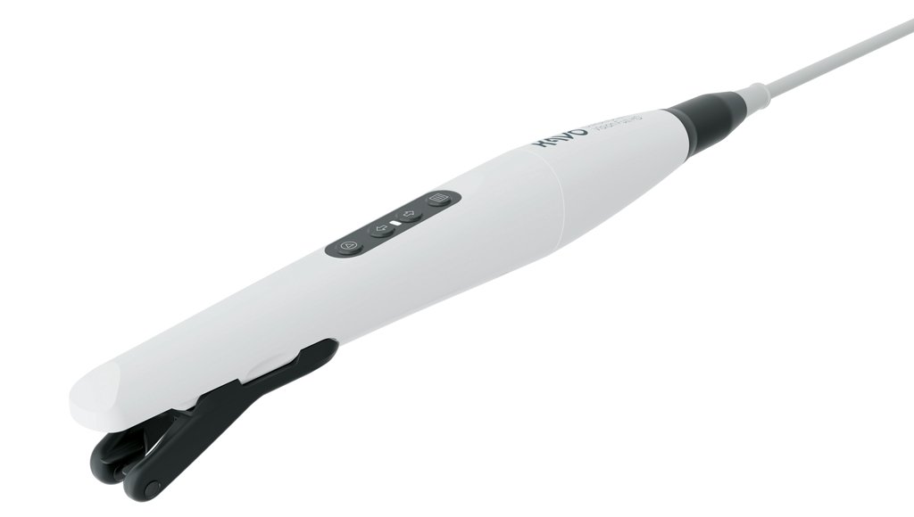
Practical design and seamless integration into existing workflows are critical factors when evaluating any new piece of dental equipment. The KaVo DIAGNOcam Vision Full HD addresses both areas with a design that prioritizes ergonomics and compatibility.
Physical Design and Ergonomics
The handpiece is lightweight and features ergonomically positioned controls directly on the camera body, allowing the operator to switch modes and capture images without removing their hands from the device. Integrated LED status indicators provide clear visual feedback during operation, confirming the active mode and image capture status. The compact form factor fits comfortably into existing operatory setups.
Patented Single-Tip Design
One of the practical advantages KaVo emphasizes is the patented single-tip design. The same tip attachment works across all three imaging modes, eliminating the need to change attachments between transillumination, fluorescence, and intraoral photography. This saves time and reduces workflow interruptions. The flexible tip arms adapt to different tooth sizes, providing consistent light delivery across the dentition. The tip can be cleaned and sterilized between patients, and replacement tips are available as consumables.
Software Compatibility
The DIAGNOcam Vision Full HD integrates with KaVo’s DTX Studio platform and connects to most third-party dental imaging and practice management software through a TWAIN driver. This broad compatibility means the device can typically be added to an existing digital workflow without requiring a software overhaul. Images are saved directly into the patient’s digital chart and can be displayed side by side across all three modes, which is especially effective for chairside patient communication.
Clinical Performance and Professional Feedback
Independent clinical evaluations and extended hands-on testing provide useful context for dental professionals considering a purchase. The feedback from clinicians who have used the KaVo DIAGNOcam Vision Full HD in daily practice has been consistently favorable.
Real-World Testing Results
Dr. John Flucke, Chief Clinical Editor and Technology Editor of Dental Products Report, tested the device for approximately four months in clinical practice. He reported that the unit is well-designed, well-balanced, and produces remarkable images (Flucke, 2024). He highlighted the autofocus feature as particularly impressive, noting that it keeps images sharp regardless of distance, including for extraoral shots. He found that an experienced user can complete a full-mouth imaging scan in under two minutes and stated that the device is highly recommended for any office looking to improve diagnostic and communication capabilities.
The Catapult Product Evaluation Team also assessed the device under real clinical conditions. More than 85% of survey respondents remarked positively on early detection capabilities and the ability to identify treatment opportunities, particularly the capacity to reveal enamel changes that were not visible on radiographs. The team awarded the DIAGNOcam the Catapult Vote of Confidence, noting that its imaging approach complements radiographs and AI-assisted diagnostics rather than competing with them (Catapult Education, 2024).
Impact on Patient Communication and Case Acceptance
One of the most frequently cited advantages is the device’s impact on patient communication. Research suggests that approximately 90% of human perception is visual, yet traditional diagnostic methods often require dentists to communicate findings verbally. The DIAGNOcam Vision Full HD changes this dynamic. With transillumination images displayed on a chairside monitor, patients can see the dark shadows of carious lesions for themselves. Clinicians consistently report that patients understand transillumination images more readily than radiographs, leading to improved case acceptance and more informed consent conversations (Flucke, DPR, 2024).
Value for Radiation-Averse Patients
A growing number of patients express concern about radiation exposure from dental radiographs, particularly parents of young children and pregnant patients. The DIAGNOcam Vision Full HD provides a radiation-free diagnostic option that can serve as a complement to, or in appropriate clinical scenarios a substitute for, bitewing radiographs. Clinical research supports the reliability of near-infrared transillumination for caries detection in both adults and children, including studies published in the Nigerian Journal of Clinical Practice (Alamoudi et al., 2019) and Biotechnology & Biotechnological Equipment (Marinova-Takorova et al., 2016), making it a scientifically justified option rather than merely a patient-pleasing compromise.
KaVo DIAGNOcam Vision Full HD vs. Traditional Diagnostic Methods
Understanding where the DIAGNOcam Vision Full HD fits relative to established diagnostic methods is essential for making an informed purchasing decision. The following comparisons outline the key differences and overlaps.
Compared to Bitewing Radiographs
Bitewing radiographs remain the gold standard for interproximal caries detection. However, clinical studies demonstrate that near-infrared transillumination achieves comparable accuracy for dentin-level lesions and superior sensitivity for initial enamel lesions. A systematic review and meta-analysis published in the Journal of Dentistry confirmed that NILT achieved diagnostic performance on par with bitewings for proximal caries detection (Defined et al., 2020). The DIAGNOcam also provides information about the buccolingual extent of lesions, eliminates radiation exposure, can be repeated without dosimetric concerns, and delivers instant results. Bitewing radiographs, on the other hand, provide information about bone levels, periapical conditions, and overall dental anatomy that transillumination cannot replicate. The most effective approach for most practices is to use both methods together.
Compared to Standard Intraoral Cameras
A standalone intraoral camera captures visible-light photographs that show surface conditions but cannot reveal subsurface pathology. The DIAGNOcam Vision Full HD adds transillumination for structural assessment and fluorescence for biological confirmation, making it a true diagnostic tool rather than merely a documentation device. The continuous autofocus also represents a practical improvement over fixed-focus lenses found in most competing cameras.
Compared to Other Caries Detection Devices
Laser fluorescence devices such as the DIAGNOdent provide numerical readings at a specific point but do not produce spatial images. The DIAGNOcam Vision Full HD provides full visual maps of lesion distribution and integrates caries detection with standard imaging, offering a more intuitive and comprehensive diagnostic experience. For practices already using intraoral scanners for restorative workflows, the DIAGNOcam adds a diagnostic dimension that scanning alone does not provide.
Who Should Consider the KaVo DIAGNOcam Vision Full HD?
The device is well suited for several practice profiles. Understanding whether it aligns with a specific clinical philosophy and patient base can help determine if the investment makes sense.
Prevention-focused practices. Clinics that emphasize early detection, remineralization therapy, and minimally invasive treatment benefit most from the transillumination and fluorescence capabilities.
Pediatric practices. The radiation-free nature of the device is especially valuable for pediatric populations, where minimizing radiation exposure is a priority.
Practices investing in patient communication. The visual communication power of transillumination images, which patients find easier to understand than radiographs, makes it a high-value investment for improving case acceptance.
High-volume practices. The one-click, three-image capture and sub-two-minute full-mouth scan capability makes multi-modal imaging practical even in a busy schedule.
Practices serving radiation-averse patients. For clinicians who regularly encounter patients declining radiographs, the DIAGNOcam provides a scientifically validated alternative for caries screening.
Bottom Line
The KaVo DIAGNOcam Vision Full HD earns a strong recommendation for dental practices that prioritize early caries detection, visual patient communication, and minimally invasive treatment philosophies. The 3-in-1 design consolidates what previously required multiple separate devices into a single, efficient workflow, and the underlying near-infrared transillumination technology has a solid evidence base across multiple peer-reviewed publications. Clinicians who have tested the device in daily practice consistently report that it improves diagnostic confidence, enhances patient understanding, and integrates smoothly into existing digital workflows.
From a return-on-investment perspective, the device addresses several revenue and efficiency drivers simultaneously. Improved case acceptance through visual patient education, earlier detection of lesions that might otherwise progress to more complex and costly restorations, and the ability to document findings with three image types in under one second all contribute to a measurable impact on practice productivity. For offices that see a high volume of hygiene patients or that operate within a prevention-focused care model, the diagnostic information captured during routine visits can directly inform treatment recommendations and support more structured follow-up protocols.
The device does require a meaningful upfront investment, and practices should set realistic expectations for the learning curve involved in interpreting transillumination and fluorescence images. It does not replace radiographic imaging for bone-level assessment, periapical diagnostics, or evaluating the full range of anatomical structures. However, for what it does deliver, which is radiation-free caries detection with clinical accuracy comparable to bitewing radiographs, combined with HD photography and bacterial fluorescence detection, the KaVo DIAGNOcam Vision Full HD occupies a unique and well-defended position in the market.
Verdict
<p>The KaVo DIAGNOcam Vision Full HD represents a meaningful advancement in chairside diagnostic technology. The 3-in-1 design, combining transillumination, fluorescence, and HD intraoral photography in a single handpiece with one-click capture, eliminates the workflow friction that previously made multi-modal imaging impractical for routine clinical use. The continuous autofocus delivers consistently sharp images without manual adjustment, translating directly into time saved across a full day of patient care.</p><p>The clinical evidence supporting near-infrared transillumination for caries detection is robust and growing, with multiple peer-reviewed studies confirming its reliability for both enamel and dentin lesions. The ability to detect caries at earlier stages than conventional radiography aligns with the profession’s ongoing shift toward minimally invasive, prevention-oriented approaches. For practices that invest in patient education and visual communication, the DIAGNOcam Vision Full HD is likely to pay for itself through improved case acceptance.</p><p>That said, the device is a complement to radiographic imaging, not a replacement. Practices should plan to integrate it alongside X-rays rather than substitute one for the other. The initial investment is significant, and interpreting transillumination and fluorescence images does require a learning period. For practices that make that commitment, the diagnostic, communication, and workflow advantages are substantial enough to warrant serious consideration.</p>
Frequently Asked Questions
What is the KaVo DIAGNOcam Vision Full HD used for?
The KaVo DIAGNOcam Vision Full HD is used for dental diagnostics, patient communication, and clinical documentation. Its primary clinical application is caries detection using near-infrared transillumination, which identifies occlusal, interproximal, and secondary caries as well as cracks and fractures without radiation. It also provides fluorescence imaging for detecting active bacterial metabolism and a high-resolution intraoral camera for standard clinical photography.
How does the DIAGNOcam detect cavities without X-rays?
The device uses near-infrared light at 780 nm to illuminate the tooth from the cervical region. Enamel is highly transparent to near-infrared light, so the tooth acts as a light conductor. A digital sensor captures the transmitted light, and areas of demineralization, decay, or structural compromise scatter the light differently, appearing as darker shadows in the grayscale image. Clinical studies have validated the effectiveness of this technology for caries detection at both enamel and dentin levels.
Is the DIAGNOcam Vision Full HD as accurate as dental X-rays?
Clinical research shows that near-infrared transillumination achieves comparable diagnostic accuracy to bitewing radiography for dentin caries and may be more sensitive for initial enamel lesions. An in vitro study found a stronger correlation between DIAGNOcam images and micro-CT (0.82) than between digital radiography and micro-CT (0.73). However, the device has limitations in assessing caries depth in dentin and cannot evaluate bone levels or periapical conditions. It works best as a complement to radiographic imaging.
Can the DIAGNOcam replace bitewing X-rays?
In certain clinical scenarios, such as initial caries screening, routine monitoring, and patient education, the DIAGNOcam can serve as an alternative to bitewing radiographs, particularly when radiation avoidance is clinically appropriate. However, for comprehensive diagnosis including bone level assessment and periapical evaluation, radiographs remain necessary. Most experts recommend using the DIAGNOcam alongside radiographic imaging rather than as a standalone replacement.
What software works with the KaVo DIAGNOcam Vision Full HD?
The device integrates with KaVo’s DTX Studio platform and connects to most third-party dental imaging and practice management software through a TWAIN driver interface. This broad compatibility allows it to fit into most existing digital workflows without requiring a software change.
How long does a full-mouth scan take?
According to clinicians who have tested the device extensively, an experienced operator can complete a full-mouth imaging scan in approximately two minutes. Each individual capture takes less than one second and produces all three image types simultaneously.
Does the device require special training?
The device is designed for intuitive operation, but interpreting transillumination and fluorescence images requires some training and clinical experience. KaVo offers educational resources including CE webinars and personalized virtual sessions with product managers. Most practitioners report developing comfort within the first few weeks of regular use.
Is the DIAGNOcam Vision Full HD safe for patients?
Yes. The device uses non-ionizing near-infrared light and visible-spectrum LEDs with no radiation exposure. The imaging process is painless, non-invasive, and can be repeated as often as clinically necessary without safety concerns, making it suitable for children, pregnant women, and patients requiring frequent monitoring.
Can the DIAGNOcam detect cracks in teeth?
Yes. Transillumination is particularly effective for identifying cracks and fracture lines. When near-infrared light passes through a crack, the disruption in the light path makes the fracture visible as a dark line in the image. Clinicians who have evaluated the device specifically noted its ability to reveal fractures missed by other diagnostic methods.
Does the device work on teeth with existing restorations?
The intraoral camera and fluorescence modes work normally on restored teeth. For transillumination, effectiveness depends on the restoration material. Composite restorations generally allow sufficient light transmission for useful diagnostic images, while large metallic restorations such as amalgam fillings or metal crowns may block near-infrared light and limit the transillumination image for those teeth.