Dental Implant Treatment Planning for Dental Professionals and Patients
A successful dental implant starts long before the surgeon picks up a handpiece. The foundation of every predictable implant outcome is a thorough, well-structured dental implant...
Written by Marcus Hale
Read time: 8 min read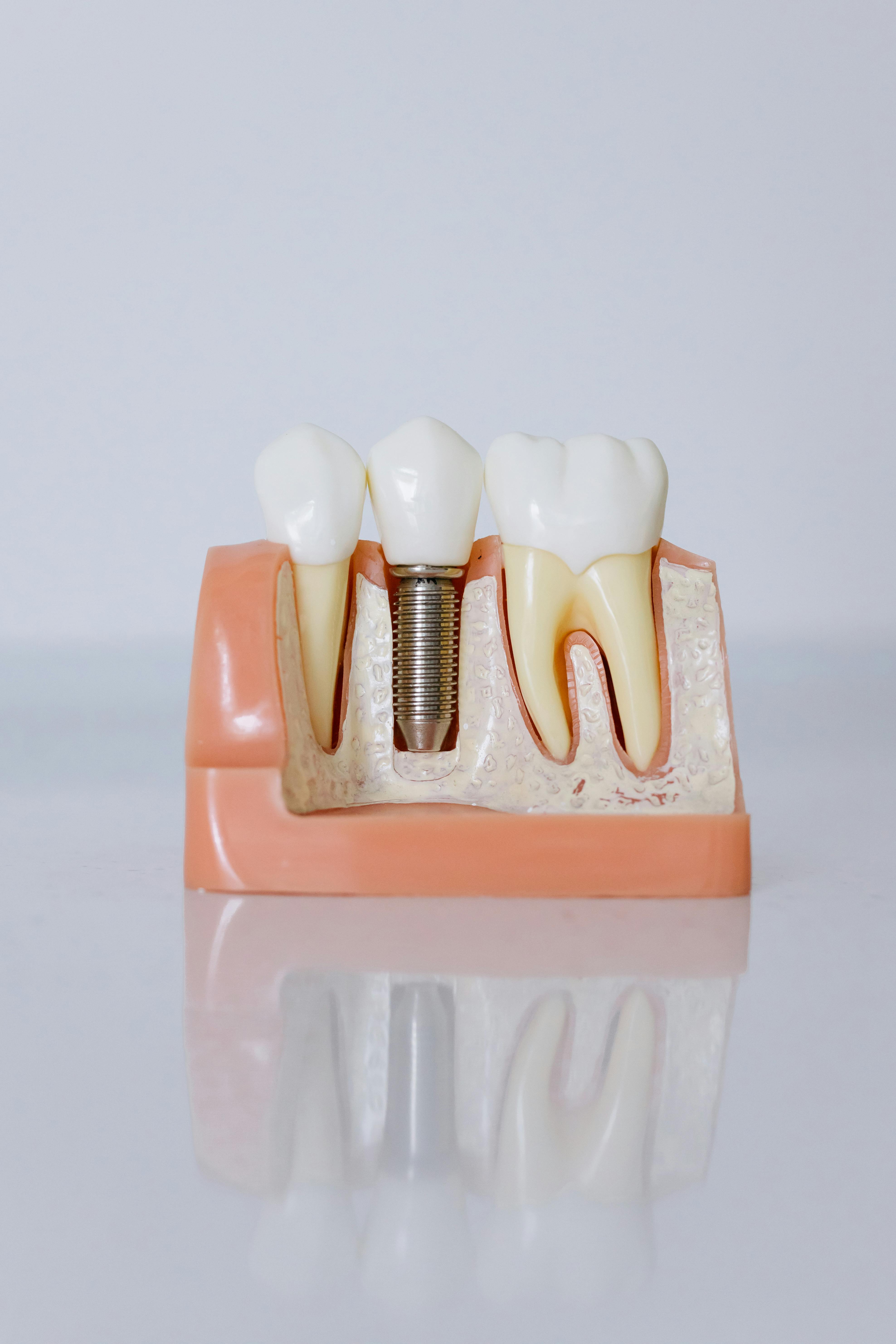
A successful dental implant starts long before the surgeon picks up a handpiece. The foundation of every predictable implant outcome is a thorough, well-structured dental implant treatment plan that accounts for the patient’s anatomy, health history, prosthetic goals, and long-term maintenance needs. Without this roadmap, the risk of complications rises significantly.
This guide covers every stage of dental implant treatment planning, from initial diagnostics and clinical assessment to modern planning software and AI-assisted workflows. It is written for two audiences: clinicians looking to refine their implant planning protocols, and patients who want to understand what a dental implant treatment plan should include and what to expect throughout the process.
Here is what this article covers:
What a dental implant treatment plan is and why it matters
The key components every plan should include
A step-by-step walkthrough of the planning process
How dental implant treatment planning software is changing clinical workflows
The role of AI in dental implant treatment planning
A realistic example of what a completed treatment plan looks like
What Is a Dental Implants Treatment Plan?
A dental implant treatment plan is a structured, written document that outlines every clinical and prosthetic step required to replace one or more missing teeth with implant-supported restorations. It goes well beyond a simple list of procedures or a cost estimate. A comprehensive plan integrates diagnostic findings, surgical strategy, prosthetic design, a realistic timeline, and a clear explanation of risks and alternatives.
For clinicians, the treatment plan serves as the project’s blueprint. It coordinates the work of the restorative dentist, the surgeon (if separate), the dental laboratory, and any additional specialists involved in the case. For patients, it provides transparency, helping them understand what will happen at each stage, how long the process will take, and what costs to anticipate.
Research consistently supports the value of thorough planning. A 2024 meta-analysis published in Clinical Oral Investigations examined 20-year implant survival data and found that long-term success depends heavily on case selection, precise surgical placement, and structured follow-up, all of which stem from robust treatment planning. The study reported 20-year survival rates between 78% and 92%, depending on the methodology used, reinforcing the point that planning quality directly influences how long implants last.
A dental implant treatment plan is different from a treatment estimate. An estimate tells patients the cost. A plan tells them the why, how, and when of every procedure, and it gives the clinical team a shared reference point for decision-making throughout the case.
Who Benefits From Dental Implant Treatment Planning?
Every patient receiving a dental implant benefits from structured treatment planning, regardless of case complexity. The scope and detail of the plan naturally scale with the clinical demands of the case, but even a straightforward single-tooth replacement deserves a written plan that documents diagnostic findings, implant selection, and prosthetic intent.
Cases that demand especially thorough dental implant treatment planning include:
Single-tooth replacements in the aesthetic zone, where the implant position directly affects the final smile
Multiple adjacent implants requiring coordinated spacing and load distribution
Full-arch rehabilitation, such as All-on-4 or All-on-6 protocols, where four to six implants support an entire arch of teeth
Cases involving bone augmentation procedures such as sinus lifts, ridge augmentation, or socket grafting
Medically complex patients with diabetes, osteoporosis, a history of bisphosphonate use, or uncontrolled periodontal disease
From the clinician’s perspective, a detailed plan helps coordinate multidisciplinary teams. When one practitioner extracts the tooth, another places the implant, and a third designs the restoration, the treatment plan acts as the shared communication document that keeps everyone aligned.
Patients benefit from receiving a written plan because it allows them to ask informed questions, compare options, seek second opinions, and budget for the full scope of treatment. A dental treatment plan template can help practices standardize this document across their patient base.
Key Components of a Dental Implant Treatment Plan
A well-constructed dental implant treatment plan contains several interconnected elements. Each component builds on the one before it, moving from diagnosis through to the final restoration. Omitting any of these steps introduces unnecessary risk.
Patient Assessment and Medical History
Planning begins before any imaging is taken. A thorough review of the patient’s medical history identifies systemic factors that may influence implant healing or increase the risk of failure. These factors include uncontrolled diabetes, autoimmune conditions, radiation therapy to the head and neck, smoking, and certain medications such as bisphosphonates or anticoagulants.
Patients should also be assessed for parafunctional habits like bruxism, which places excessive load on implant restorations and may require protective measures such as a night guard.
Clinical Examination
The intraoral clinical examination evaluates the soft and hard tissues at the proposed implant site. Clinicians assess the width and height of the edentulous ridge, the quality of the keratinized tissue, the occlusal relationship, and the condition of adjacent teeth. This examination also identifies active periodontal disease, which must be stabilized before implant placement can proceed.
Diagnostic Imaging
Two-dimensional radiographs, such as panoramic films, provide an initial overview, but three-dimensional cone beam computed tomography (CBCT) has become the standard of care for dental implant treatment planning. CBCT scans reveal the precise dimensions of available bone, the location of vital structures such as the inferior alveolar nerve and the maxillary sinus floor, and any pathology that may not be visible on conventional imaging.
Radiation doses from modern CBCT units are considerably lower than those from medical CT scanners, making them appropriate for routine implant planning. For clinicians evaluating CBCT scanners for their practice, image resolution, field of view flexibility, and software integration are the most important selection criteria.
Bone Quality and Quantity Assessment
CBCT data allows clinicians to measure ridge height, width, and angulation at the planned implant site. Bone density can be estimated through Hounsfield unit values, helping the surgeon anticipate primary stability during placement. When bone volume is insufficient, augmentation procedures such as guided bone regeneration, sinus floor elevation, or block grafting must be incorporated into the treatment plan.
The proximity of the implant site to vital anatomical structures also requires careful evaluation. In the mandible, the inferior alveolar nerve canal sets the lower boundary for safe implant placement, with most practitioners maintaining a safety margin of at least 2 mm. In the posterior maxilla, the sinus floor determines available bone height, and cases with less than 5–6 mm of residual bone typically require a sinus lift before implant placement. Thorough bone assessment at the planning stage prevents intraoperative surprises and allows the team to have all necessary grafting materials and instruments prepared in advance.
Prosthetic-Driven Planning
Modern implant treatment planning works backward from the desired prosthetic result. Rather than placing the implant where bone is most available and then adapting the restoration, clinicians now determine the ideal tooth position first and then plan the implant to support it. Diagnostic wax-ups, digital smile design, and virtual tooth set-ups are all used to establish the target restoration before surgery.
This approach, often called “prosthetic-driven” or “restoratively driven” planning, produces better aesthetic outcomes and more favorable loading mechanics. It also highlights cases where bone augmentation will be needed to achieve the ideal implant position.
Timeline and Phasing
Dental implant treatment rarely happens in a single appointment. The plan should clearly outline the sequence and timing of each phase: tooth extraction (if needed), bone grafting (if needed), implant placement, the healing and osseointegration period (typically three to six months), abutment connection, and final restoration delivery.
Some cases allow for immediate implant placement at the time of extraction, and some allow for immediate loading with a provisional restoration on the same day. The treatment plan should specify the chosen protocol and explain the rationale behind it.
Cost Estimation and Informed Consent
Patients should receive a clear, itemized cost breakdown before treatment begins. The plan should list the fees for each procedure (imaging, surgery, bone grafting, abutment, restoration) along with any laboratory costs. It should also document the informed consent discussion, including the risks of treatment, the consequences of no treatment, and any alternative options such as bridges or removable dentures.
Practices looking to streamline this process can use a dental treatment plan generator to produce consistent, professional documents that cover all the required elements.
The Dental Implant Treatment Planning Process: Step by Step
Understanding the treatment planning workflow helps both clinicians and patients appreciate how each stage contributes to the final result. The following sequence represents a typical implant planning process from the first visit to plan delivery.
Step #1: Initial consultation and goal setting. The clinician meets with the patient to discuss their concerns, expectations, and goals. This conversation establishes the desired outcome and begins the process of managing expectations around timelines, aesthetics, and cost.
Step #2: Records gathering. Clinical photographs, intraoral scans or conventional impressions, and radiographic images (panoramic and CBCT) are captured. These records form the diagnostic foundation of the plan.
Step #3: Diagnostic analysis and case design. The clinician reviews the imaging data, assesses bone and soft tissue conditions, and uses planning software to virtually position the implant in the ideal prosthetic location. A digital wax-up or smile design may be created at this stage.
Step #4: Multidisciplinary review. In complex cases, the surgeon, restorative dentist, and laboratory technician review the proposed plan together. This review ensures alignment between the surgical approach and the prosthetic vision. Digital workflows make this collaboration easier, as CBCT data and intraoral scans can be shared and merged in planning software.
Step #5: Plan presentation to the patient. The completed treatment plan is presented to the patient with visual aids, cost estimates, and a clear timeline. Software-generated 3D renderings can help patients visualize the planned outcome and understand the rationale behind each procedure.
Step #6: Informed consent and scheduling. Once the patient agrees to the plan, informed consent is documented, and appointments are scheduled. If a surgical guide will be used, it is ordered or designed at this stage.
For a deeper look at how to structure treatment plans across all dental disciplines, see this clinical guide to preparing a dental treatment plan.
Dental Implant Treatment Planning Software: Tools Shaping Modern Workflows
Dental implant treatment planning software has fundamentally changed how clinicians approach implant cases. These platforms integrate CBCT data with intraoral scans, allow virtual implant placement in three dimensions, simulate the prosthetic outcome, and generate files for surgical guide fabrication. The result is a more predictable surgical outcome and a more efficient workflow from planning through to restoration.
Most dental implant treatment planning software falls into one of the following categories:
Standalone implant planning platforms. These are dedicated tools designed specifically for implant surgery planning. Examples include coDiagnostiX (Dental Wings), DTX Studio Implant (Nobel Biocare), Blue Sky Plan (Blue Sky Bio), and 3Shape Implant Studio. They typically accept DICOM files from any CBCT unit and STL files from most intraoral scanners.
Integrated CAD/CAM ecosystems. Some manufacturers offer planning modules built into broader digital dentistry platforms. Planmeca Romexis, for example, combines CBCT viewing, implant planning, and surgical guide design within a single software environment.
Cloud-based solutions. Newer platforms operate in the cloud, enabling remote collaboration between the clinician, the laboratory, and specialists. This model simplifies case sharing and eliminates the need for local software installations.
When evaluating dental implant treatment planning software, clinicians should consider the following factors:
DICOM and STL compatibility to ensure the software works with their existing imaging equipment and intraoral scanners
Open vs. closed architecture, which determines whether surgical guides and prosthetic files can be exported to third-party providers
Implant library breadth, as the software should support the implant systems the practice uses
Surgical guide design and export capabilities, including compatibility with in-office 3D printers
The learning curve, including available training and support resources
Pricing model, comparing subscription-based access against one-time license fees
A narrative review published in Bioengineering examined digital technologies in implantology and confirmed that software-guided planning significantly reduces angular and linear deviations compared to freehand techniques, translating into more accurate implant placement and fewer complications.
Planning software also plays an important patient-facing role. Three-dimensional visualizations and simulated outcomes make it easier for patients to understand their dental implant treatment plan, which improves case acceptance rates and reduces anxiety.
AI Dental Implant Treatment Planning: What’s Real and What’s Next
Artificial intelligence is beginning to play a meaningful role in dental implant treatment planning, though it is important to distinguish between capabilities that are already in clinical use and those that remain experimental. AI dental implant treatment planning is currently focused on automating time-consuming diagnostic tasks and providing decision support, not on replacing clinical judgment.
The most established AI applications in implant planning include:
Automated CBCT segmentation. AI algorithms can automatically identify and trace anatomical structures on CBCT scans, including the inferior alveolar nerve canal, maxillary sinus boundaries, and individual tooth roots. This saves significant chairside time and reduces the risk of human error in landmark identification.
Bone density mapping. Machine learning models can analyze CBCT data to classify bone quality across the implant site, helping clinicians anticipate primary stability and choose appropriate drilling protocols.
AI-suggested implant positioning. Some platforms now offer algorithm-generated implant placement suggestions that account for available bone, proximity to vital structures, and prosthetic requirements. The clinician reviews and adjusts these suggestions before finalizing the plan.
Diagnostic screening. AI-powered tools such as Overjet and Pearl analyze radiographic images to flag potential pathology, periapical lesions, and bone loss patterns, providing a second-opinion layer before treatment planning begins.
These tools offer tangible benefits. Automated segmentation reduces CBCT analysis time from 30–45 minutes to a few minutes. AI-suggested positioning gives less experienced clinicians a validated starting point for implant placement. Diagnostic screening adds a safety net that catches findings a busy clinician might overlook.
However, limitations remain. Most AI tools function as decision-support systems, meaning the clinician retains full responsibility for the final plan. Training data biases can affect accuracy in certain anatomical situations. Regulatory approvals vary across markets, and not all AI features have undergone the level of clinical validation that clinicians should expect before trusting automated outputs.
Looking ahead, AI dental implant treatment planning is likely to integrate with robotic-assisted surgery, real-time intraoperative navigation, and predictive outcome modeling that draws on large patient datasets. These developments could make implant treatment planning more personalized and more accessible, particularly in regions where specialist expertise is limited.
For clinicians considering AI-enhanced tools, the practical advice is to start with platforms that offer validated diagnostic features such as nerve tracing and bone segmentation, and to treat AI-generated implant placement suggestions as a starting point rather than a final answer. As more clinical validation studies are published, the evidence base for these tools will continue to strengthen.
Example: What a Dental Implant Treatment Plan Looks Like
Patients searching for a dental implant treatment plan example often want to see what the document actually contains. The following is a simplified, fictional example based on a common clinical scenario. Real treatment plans will vary depending on the case, the clinician, and the practice.
Our dental treatment plan generator, assisted by an AI trained on anonymized real-life cases, has generated this exemplary dental implant treatment plan.
Patient summary. Female patient aged 18–35, no significant medical history noted. Missing or non-restorable maxillary right canine (tooth #6). Patient requests a fixed, implant-supported replacement to restore aesthetics within the "social six" and re-establish canine guidance during lateral excursions.
Diagnostic findings. CBCT scan to be completed to evaluate bone density, volume, and 3D positioning at the #6 site. Assessment of the alveolar ridge for potential localized atrophy and evaluation of keratinized mucosa width. The proximity to the maxillary sinus and the lateral nasal wall needs to be confirmed. Adjacent teeth (#5 and #7) are healthy. High smile line noted, requiring precise soft tissue management. Facial growth completion to be verified if the patient is at the younger end of the age range (serial cephalometric tracing if necessary). Possible history of bruxism to be assessed.
Proposed treatment:
Extraction of tooth #6 (if still present) using an atraumatic technique with socket preservation via bone graft and collagen membrane
Provision of a temporary aesthetic solution (Essix retainer or bonded Maryland bridge)
Prophylaxis, oral hygiene instruction, and management of any active caries or periodontal disease
CBCT analysis and 3D implant planning
Placement of one endosseous implant (titanium or zirconia) at the #6 site using a restoratively driven surgical guide
3–6 months of undisturbed osseointegration
Second-stage surgery for healing, abutment placement (if submerged protocol used)
Digital or conventional impression
Delivery of a custom CAD/CAM zirconia abutment with a screw-retained or cement-retained porcelain-fused-to-zirconia (PFZ) crown
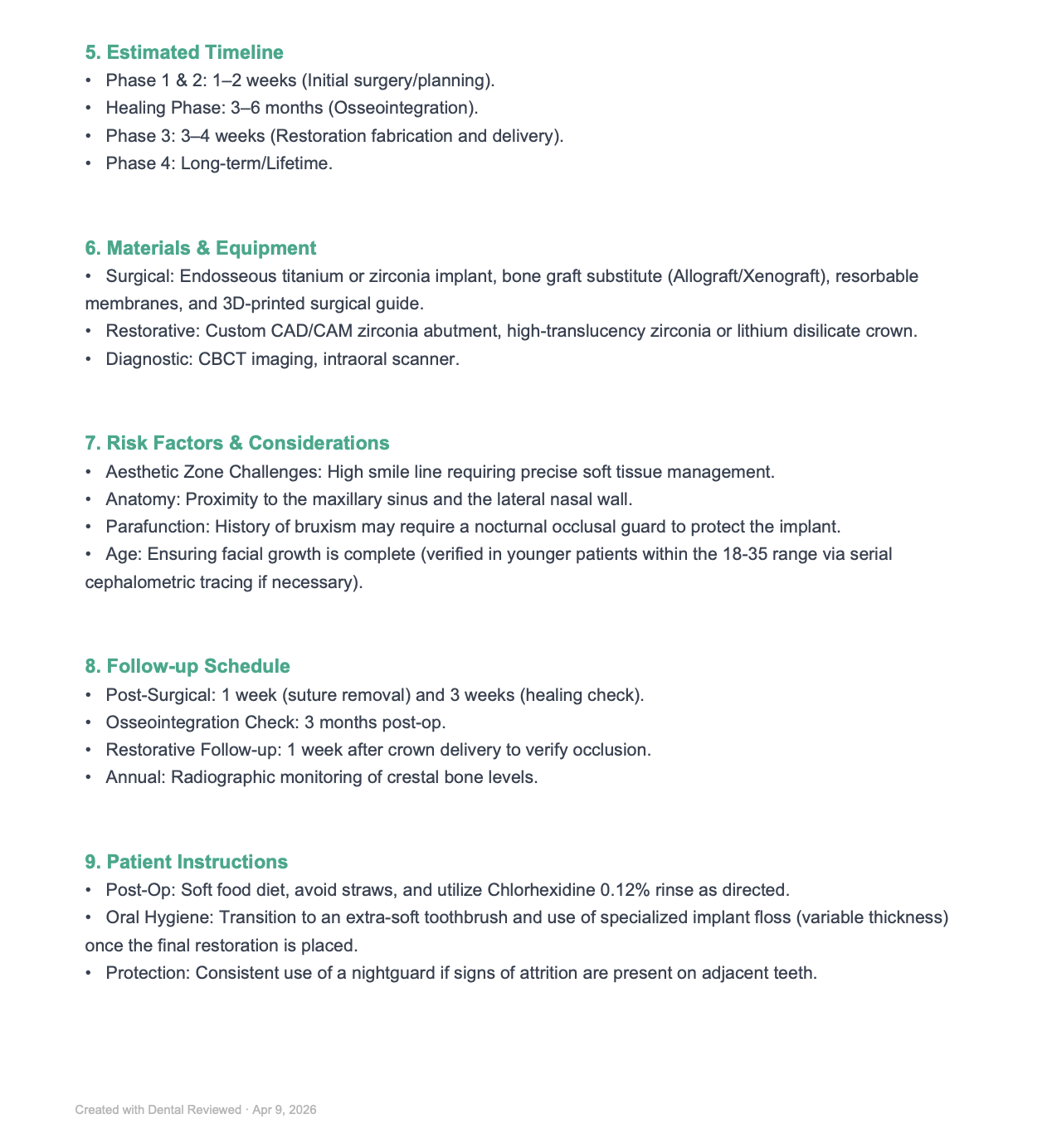
Timeline:
Weeks 0–2. Extraction, socket preservation, temporary prosthesis, and initial planning
Months 1–6. Osseointegration healing period
Months 6–7. Final restoration fabrication and crown delivery
Ongoing. Integration into a 6-month professional hygiene recall
Materials and equipment:
Surgical. Endosseous titanium or zirconia implant, bone graft substitute (allograft/xenograft), resorbable collagen membrane, 3D-printed surgical guide
Restorative. Custom CAD/CAM zirconia abutment, high-translucency zirconia or lithium disilicate crown
Diagnostic. CBCT imaging, intraoral scanner
Risks discussed. Implant failure, infection, aesthetic zone complications due to high smile line, proximity to the maxillary sinus and lateral nasal wall, and peri-implant tissue instability. Bruxism may require a nocturnal occlusal guard to protect the implant and adjacent teeth. Alternative options to be discussed with the patient.
Follow-up schedule:
1 week post-surgery. Suture removal
3 weeks post-surgery. healing check
3 months post-op. Osseointegration verification
1 week after crown delivery. Occlusion and proximal contact check, baseline peri-implant radiographs
Annually. Radiographic monitoring of crestal bone levels
Post-operative instructions. Soft food diet, no straws, chlorhexidine 0.12% rinse as directed. Transition to an extra-soft toothbrush once healed. Specialized implant floss (variable thickness) once the final restoration is placed. Consistent nightguard use if signs of attrition are present.
Here's how the dental implant treatment plan looks in general:
Keep in mind that this example is based on a fictional treatment plan generated for illustration. Actual plans will differ based on clinical findings, the implant system used, patient-specific anatomy, and practitioner protocols. Every treatment plan should be reviewed by a dental professional before implementation, taking into account the individual patient's condition, comorbidities, drug interactions, and other clinically relevant factors.
Common Challenges in Dental Implant Treatment Planning
Even experienced clinicians encounter situations that complicate the planning process. Recognizing these challenges early allows for better preparation and more realistic expectations for both the treatment team and the patient.
Insufficient bone volume. Bone resorption following tooth loss is one of the most common obstacles. When the remaining ridge is too narrow or too short to accommodate an implant, grafting procedures must be incorporated into the plan. This adds cost, extends the treatment timeline, and introduces additional healing variables.
Proximity to vital structures. In the posterior mandible, the inferior alveolar nerve limits how deep an implant can be placed. In the posterior maxilla, the maxillary sinus may encroach on available bone height. Both situations require careful CBCT analysis and, in some cases, alternative approaches such as shorter implants, angled placement, or sinus lift procedures.
Managing patient expectations. Patients sometimes expect implant treatment to be faster, cheaper, or less invasive than it actually is. The treatment plan is the right place to set realistic expectations about healing times (often 3–6 months for osseointegration alone), the possibility of staged procedures, and the aesthetic limitations that may apply in cases with significant bone or soft tissue loss.
Multidisciplinary coordination. When the referring dentist, the surgeon, and the prosthodontist work in different practices, communication gaps can arise. Digital workflows help bridge this gap, as planning files, CBCT data, and scan files can be shared electronically. Practices investing in digital scanners and compatible planning software find that coordination becomes significantly smoother.
Plans that go stale. Treatment plans have a shelf life. If a patient delays treatment for months or years, bone conditions may change, adjacent teeth may shift, and the original plan may no longer be valid. Re-imaging and re-planning are necessary in these cases, and patients should be informed that significant delays may result in additional costs.
Tips for Patients Reviewing a Dental Implant Treatment Plan
Patients who take an active role in understanding their treatment plan are better prepared for the process and more satisfied with the outcome. The following questions can help patients evaluate the quality and completeness of the plan they receive.
Questions to ask your dentist:
What imaging was used to plan my implant, and was a CBCT scan taken?
Why was this particular implant system chosen for my case?
What are the alternatives to implant treatment, and how do they compare in terms of longevity and cost?
What happens if something changes during surgery and the original plan cannot be followed?
How long will the entire process take from start to finish?
Is the cost estimate inclusive of all fees, including the laboratory and follow-up visits?
A good treatment plan should be written, detailed, and easy to understand. It should include diagnostic findings, the proposed procedures in sequence, a timeline, and a cost breakdown. If a plan feels vague or rushed, or if it was presented verbally without any written documentation, patients should consider seeking a second opinion.
Patients should also feel comfortable asking about the clinician’s experience with implant cases similar to theirs. Complex cases such as full-arch rehabilitation or cases requiring significant bone grafting should ideally be managed by practitioners with advanced training and a track record of successful outcomes.
It is also worth asking about the technology used in the planning process. Practices that use CBCT imaging, digital scanning, and dedicated implant planning software are more likely to deliver precise, predictable outcomes than those relying solely on two-dimensional radiographs and manual planning methods. The investment a practice makes in its diagnostic and planning technology often reflects its commitment to quality.
For those comparing treatment costs across providers, reviewing the available dental insurance plans can help identify which portion of implant treatment may be covered.
Bottom Line
Dental implant treatment planning is the single most important factor in achieving a successful, lasting implant outcome. A comprehensive plan protects both the clinician and the patient, ensuring that decisions are based on accurate diagnostics, clear communication, and a shared understanding of the goals and limitations of treatment.
The shift toward digital and AI-assisted workflows is accelerating. Dental implant treatment planning software now allows clinicians to simulate surgical and prosthetic outcomes with a level of precision that was not possible a decade ago. AI-powered tools are reducing the time required for diagnostic analysis and adding valuable safety checks to the planning process.
For clinicians, the message is clear: invest in robust planning workflows, stay current with digital tools, and ensure every patient receives a written, detailed treatment plan before any surgical intervention. For patients, the takeaway is equally straightforward: ask for a plan, read it carefully, and make sure it answers every question about what will happen, when, and at what cost.
The best treatment plans are living documents, updated as the case progresses and adjusted when clinical realities differ from initial expectations. A plan that accounts for contingencies, communicates clearly, and integrates the latest diagnostic technology gives both the clinical team and the patient the best possible foundation for a successful result.
This article is for informational purposes only and does not constitute medical advice. Always consult with qualified healthcare professionals for diagnosis and treatment recommendations specific to your situation.
Frequently Asked Questions
What is a dental implant treatment plan?
A dental implant treatment plan is a written clinical document that outlines the diagnostic findings, proposed surgical and prosthetic procedures, the treatment timeline, estimated costs, and risk considerations for placing one or more dental implants. It serves as the roadmap for the entire treatment process.
How long does the dental implant treatment planning process take?
The planning phase typically takes one to three appointments, depending on the complexity of the case. Simple single-tooth cases may be planned within a week of the initial consultation, while complex full-arch cases involving bone grafting can take several weeks to finalize.
Do I need a CBCT scan for dental implant planning?
CBCT imaging is now widely considered the standard of care for implant planning. It provides three-dimensional information about bone volume, density, and the location of anatomical structures that cannot be assessed with traditional two-dimensional radiographs alone.
What is the difference between dental implant treatment planning software and AI-assisted planning?
Dental implant treatment planning software provides the digital tools for virtual implant placement, surgical guide design, and prosthetic simulation. AI-assisted planning adds a layer of automation, such as auto-segmentation of CBCT scans and algorithm-generated implant position suggestions. AI enhances the software workflow, but the clinician remains in control of all final decisions.
How much does a dental implant treatment plan cost?
Costs vary depending on the complexity of the case, the imaging required, and the practice. A CBCT scan and treatment plan consultation typically ranges from £150 to £500. Some practices include the planning fee in the overall implant treatment cost, while others charge it separately.
Can I get a second opinion on my dental implant treatment plan?
Absolutely. Seeking a second opinion is a reasonable and common step, particularly for complex or costly treatment plans. A second clinician can review your imaging and records and provide an independent assessment of the proposed treatment.