What Is Guided Implantology?
The landscape of dental implant surgery has transformed dramatically over the past two decades. What once relied heavily on clinical intuition and two-dimensional radiographs has...
Written by Mantas Petraitis
Read time: 10 min read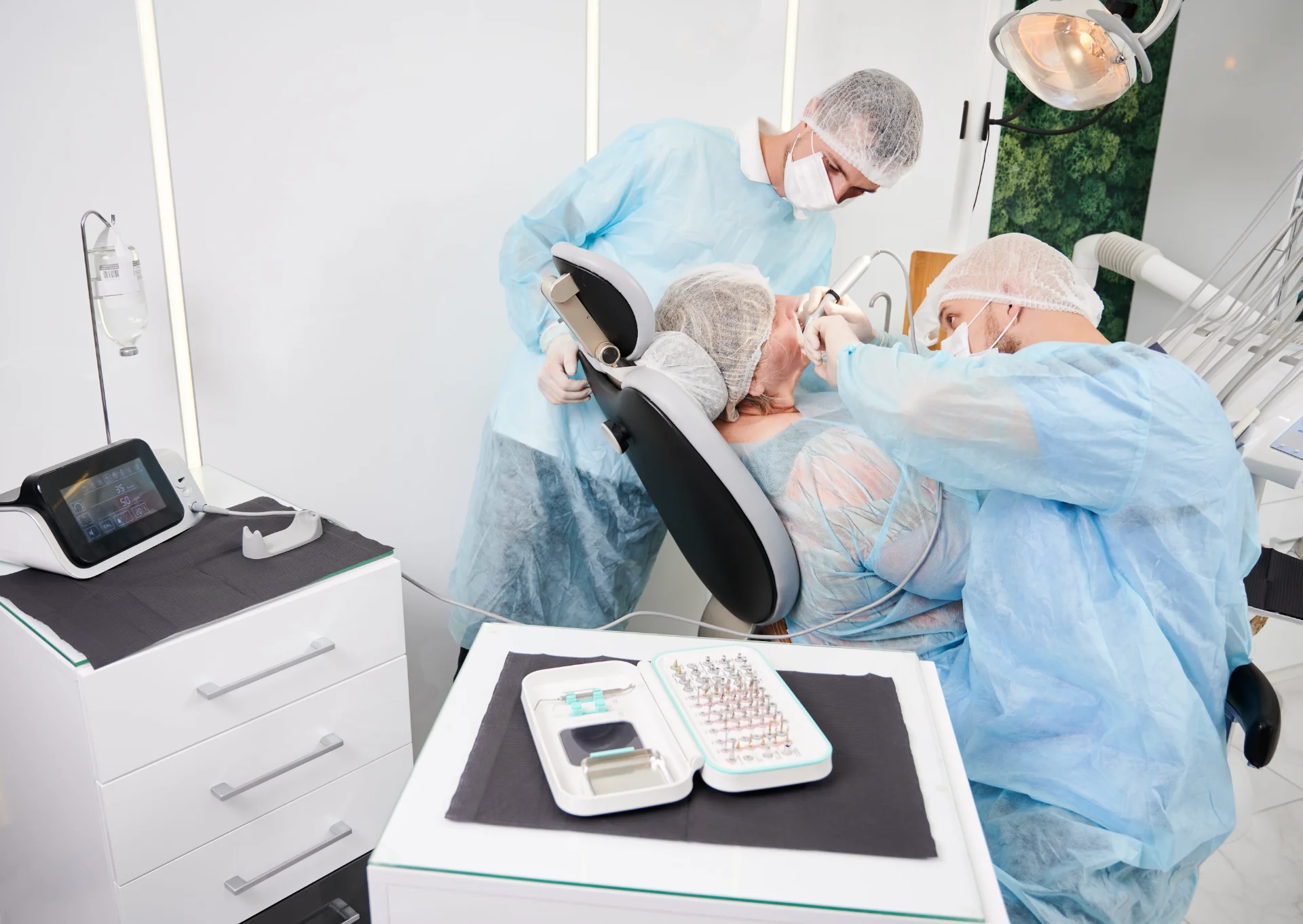
The landscape of dental implant surgery has transformed dramatically over the past two decades. What once relied heavily on clinical intuition and two-dimensional radiographs has evolved into a sophisticated digital workflow that allows clinicians to visualize, plan, and execute implant placement with unprecedented precision. Guided implantology represents this evolution, combining advanced imaging technology, virtual treatment planning, and computer-aided manufacturing to achieve optimal implant positioning.
Definition And Core Principles
Guided implantology refers to the complete process of digital planning, custom surgical guide fabrication, and implant placement using computer-aided systems paired with implant system-specific surgical kits. This approach enables clinicians to transfer a virtually planned implant position from software to the surgical site with measurable accuracy.
The fundamental principle underlying guided implantology is prosthetically-driven implant placement. Rather than placing implants where bone happens to be most abundant, clinicians plan backward from the desired final restoration. This approach considers the future prosthesis virtual envelope to determine the ideal implant position, depth, and angulation. The result is improved aesthetic outcomes with appropriate and cleanable emergence profiles.
According to research published in the British Dental Journal, inappropriate implant positioning can lead to problems ranging from simple cleaning difficulties to more serious complications such as peri-implantitis. Guided surgery addresses these concerns through meticulous preoperative planning and precise surgical execution.
Evolution From Conventional To Computer-Assisted Surgery
Traditional implant planning relied on clinical examination, study casts, and panoramic or periapical radiographs. These two-dimensional imaging techniques presented inherent limitations in assessing true bone dimensions and the spatial relationship between planned implant sites and critical anatomical structures.
The adoption of cone beam computed tomography (CBCT) overcame these limitations, allowing detailed three-dimensional evaluation of osseous topography. When combined with computer-aided design and computer-aided manufacturing (CAD/CAM) technologies, CBCT data enables surgical planning in virtual 3D environments.
Today, implant-prosthetic rehabilitations demonstrate 5-year survival rates of approximately 95% and greater than 89% after 10 years. Guided implantology aims to further improve these outcomes while reducing surgical complications such as nerve injury, sinus perforation, and lingual cortical plate perforation.
The Digital Workflow In Guided Implant Surgery
The guided implant surgery workflow follows a systematic sequence of steps, each building upon the previous to ensure accurate transfer of the virtual plan to the surgical site. Understanding each phase is essential for clinicians seeking to implement this technology in their practice.
Step #1: Data Acquisition
Accurate data acquisition forms the foundation of successful guided surgery. Errors introduced at this stage propagate through the entire workflow.
CBCT has become the standard imaging modality due to its favorable radiation dose profile. Research indicates that CBCT delivers a radiation dose of 92-118 μSv compared to 860 μSv for conventional CT. CBCT data is stored in DICOM format, allowing import into virtually all planning software systems.
Virtual dental models from intraoral optical scanning complement CBCT data, providing detailed tooth surface and soft tissue information in STL format. The alignment of CBCT with optical scan data occurs through either surface registration (matching dental surfaces directly) or fiducial marker registration using a radiographic splint. Surface registration is less time-consuming, though misalignment can occur with extensive metallic restorations.
Step #2: Virtual Treatment Planning
Virtual implant planning represents the intellectual heart of guided surgery, requiring careful consideration of both prosthetic goals and anatomical constraints.
Modern planning software allows clinicians to design virtual crowns or import prosthetic designs from CAD systems. The implant position is then determined based on this virtual restoration. Software platforms such as 3Shape Implant Studio, Planmeca Romexis, exocad exoplan, coDiagnostiX, and Nobel Clinician offer comprehensive tools for prosthetically-driven planning.
During planning, clinicians must evaluate bone quality and density, available bone volume, distance to adjacent teeth (minimum 1.5mm), distance between implants (minimum 3mm), and proximity to vital structures such as the inferior alveolar nerve (2-5mm safety margin) and maxillary sinus. The 2018 ITI consensus paper recommended maintaining a 2mm safety margin to account for potential deviations.
Step #3: Surgical Guide Design And Fabrication
Once the virtual plan is finalized, the surgical guide is designed within the planning software and manufactured using CAD/CAM technology. The design incorporates planned implant positions with appropriate sleeve systems corresponding to the specific implant manufacturer's guided surgery kit.
Stereolithographic (SLA) 3D printing has become the standard manufacturing method, with the digital design exported as an STL file and printed using biocompatible resins. In-house 3D printing has become increasingly accessible, enabling same-day guide fabrication, though guides can also be manufactured by dental laboratories or centralized production facilities.
Step #4: Surgical Execution
The surgical phase involves positioning the guide and executing the osteotomy sequence according to the manufacturer's protocol. Procedures may be pilot-guided (only initial drill trajectory controlled), partially guided (osteotomy guided but freehand implant insertion), or fully guided (both osteotomy and implant insertion through the guide). Fully guided protocols offer the highest precision but require adequate interocclusal space.
Static Vs. Dynamic Guided Surgery
Computer-assisted implant surgery encompasses two distinct approaches: static guided surgery using physical templates and dynamic navigation using real-time tracking systems.
Static Computer-Aided Implant Surgery (sCAIS)
Static guided surgery uses a prefabricated surgical template containing metal or plastic sleeves positioned according to the virtual plan. These sleeves accept system-specific drills and guide instruments that control trajectory, position, and depth. The guide is stabilized through contact with teeth, mucosa, or bone.
Advantages: lower initial investment, no additional chairside equipment, established workflow with extensive literature support, enables flapless surgery with prefabricated provisionals, and consistent results regardless of operator experience.
Limitations: no intraoperative modification possible, reduced surgical site visualization, restricted irrigation access, potential guide instability with mucosa-supported designs, and limited mouth opening can prevent access.
Dynamic Navigation Systems (dCAIS)
Dynamic navigation uses optical tracking technology to monitor the surgical handpiece position relative to the patient's anatomy. A reference device with fiducial markers is fixed to the patient, and cameras track reflective markers on both the reference device and surgical instruments. The system displays drill position superimposed on preoperative CBCT images in real-time.
According to Prof. Alessandro Pozzi's clinical guidelines, dynamic navigation is beneficial for single and partially edentulous patients, terminal dentition cases without strategic teeth, edentulous mandibles, and situations requiring intraoperative flexibility. Static templates remain preferred for anterior maxillary cases with prefabricated prosthetics and edentulous maxillae with wide palatal support.
Comparative Analysis
Research comparing both approaches demonstrates clinically acceptable accuracy for each. A prospective clinical study found that dynamic navigation achieved a mean angular deviation of 2.19° with comparable accuracy to static guides, while both methods significantly outperformed freehand placement. A meta-analysis found no statistically significant differences between static and dynamic CAIS in overall accuracy.
Types Of Surgical Guides
The support mechanism of a surgical guide significantly influences its stability, accuracy, and clinical applicability.
Tooth-Supported Guides
Tooth-supported surgical guides utilize remaining natural teeth to anchor and stabilize the template. Research consistently demonstrates that tooth-supported guides exhibit the highest accuracy due to their anchorage on hard tissue landmarks. They are indicated for single implants and partially edentulous cases, require sufficient remaining teeth for support, and should include inspection windows at interproximal areas to verify complete seating.
Mucosa-Supported Guides
Mucosa-supported guides rest directly on soft tissue and are indicated for fully edentulous patients undergoing flapless surgery. The pressure exerted during drilling, combined with mucosal resilience, may cause guide movement affecting precision. Strategies to improve accuracy include ensuring healthy, firm tissue quality, using multiple fixation pins, and accounting for anticipated mucosal compression during planning.
Bone-Supported Guides
Bone-supported guides require full-thickness flap reflection with the guide resting directly on the alveolar ridge. Fixation screws anchor the guide to the underlying bone. While bone provides a rigid support surface, these guides introduce additional surgical trauma and are reserved for cases requiring bone visualization or recontouring.
Clinical scenario | Recommended guide type |
|---|---|
Single implant with adjacent teeth | Tooth-supported |
Partially edentulous with stable teeth | Tooth-supported |
Fully edentulous, flapless protocol | Mucosa-supported with pins |
Fully edentulous, flap surgery | Bone-supported |
Accuracy Metrics And Clinical Outcomes
Understanding how accuracy is measured and what constitutes clinically acceptable deviation is essential for interpreting the literature and setting realistic expectations.
Understanding Deviation Measurements
Accuracy in guided implant surgery is quantified through several parameters: angular deviation (angle between planned and placed implant axes in degrees), coronal/platform deviation (horizontal distance at implant platform level in mm), apical deviation (horizontal distance at implant apex in mm), and depth deviation (vertical discrepancy from planned depth in mm).
Evidence-Based Accuracy Data
The most comprehensive analysis comes from a systematic review of 67 clinical trials involving 5,673 implants:
Mean coronal deviation: 1.11mm (95% CI: 1.02-1.19)
Mean apical deviation: 1.40mm (95% CI: 1.31-1.49)
Mean angular deviation: 3.51° (95% CI: 3.27-3.75)
A systematic review of 41 articles found mean distance deviation less than 2mm (most less than 1mm) and angular deviation less than 8° (most less than 5°). Research generally considers deviations of ±1-2mm clinically acceptable.
Factors Influencing Accuracy
Multiple variables affect accuracy, including CBCT image quality, registration accuracy, guide support type (tooth-supported generally most accurate), guide stability and manufacturing precision, sleeve tolerance, bone density, interocclusal space, operator experience, and edentulous span size.
Implant Survival Rates
A meta-analysis reported guided surgery's early failure rate of 2.25% versus 6.42% for freehand placement (risk ratio 0.29, p < 0.001), representing a nearly threefold reduction in risk. Long-term data demonstrate 20-year implant survival rates reaching 92%.
Clinical Indications And Case Selection
Guided implant surgery offers advantages across a spectrum of clinical scenarios, though certain situations derive particular benefit from computer-assisted approaches.
Ideal Candidates For Guided Surgery
The following characteristics favor guided surgery: anatomically challenging sites near vital structures, aesthetic zone implants requiring precise emergence profiles, immediate implant protocols, immediate loading with prefabricated prosthetics, multiple implant and full-arch cases, medically compromised patients benefiting from minimally invasive approaches, and less experienced practitioners seeking consistency.
Complex Case Applications
Guided surgery provides particular value near critical anatomical structures. Research demonstrates that ideal implant placement through guided surgery reduces complications such as nerve injury and cortical plate perforation.
For immediate implant placement, guided surgery allows precise planning relative to the socket walls. Studies indicate that flapless computer-guided immediate placement preserves soft tissue architecture and reduces marginal bone loss.
Full-arch rehabilitations demand accurate positioning for prefabricated prosthetic delivery. Navigation-guided surgery for complete arch cases has demonstrated platform deviation of 1.17mm and angular deviation of 2.19°, supporting immediate loading protocols.
Flapless Surgery Considerations
Flapless surgery offers reduced bleeding, less postoperative discomfort, faster healing, and soft tissue preservation. According to British Dental Journal research, prerequisites include adequate bone width (minimum 6-7mm), sufficient keratinized tissue, healthy mucosal quality, and accurate surgical guide fit. Contraindications include insufficient bone requiring augmentation, need for bone visualization, thin tissue biotype, and inadequate keratinized tissue. Research indicates that freehand flapless surgery without guidance increases the risk of malposition and bone dehiscence.
Contraindications And Limitations
Absolute contraindications: restricted mouth opening preventing guide seating, insufficient bone requiring pre-implant augmentation, and severe CBCT artifacts preventing accurate planning.
Relative limitations: limited interocclusal distance in posterior regions, poor bone density affecting stability, need for significant bone recontouring, and severely resorbed ridges requiring visualization.
Sources Of Error And Risk Mitigation
Guided implant surgery involves multiple sequential steps, each presenting opportunities for error accumulation.
Error Accumulation Throughout The Workflow
The precision of guided surgery depends on the interplay of errors accumulated throughout the process. Research classifies these into image acquisition errors (CBCT artifacts, patient movement, registration inaccuracies), guide-related errors (3D printing inaccuracies, sleeve tolerances, guide deformation), and surgical errors (incomplete seating, guide movement, drill deflection, inadequate irrigation).
Risk Mitigation Strategies
During image acquisition: use cotton rolls during CBCT, ensure the patient remains motionless, and verify scan quality before planning.
During planning: master one software system, verify registration accuracy, apply 2mm safety margins, and consider peer review for complex cases.
During guide fabrication: use validated biocompatible resins, verify print accuracy on models, include inspection windows, and confirm sleeve positioning.
During surgery: verify complete guide seating, use fixation pins for mucosa/bone-supported guides, maintain irrigation, follow manufacturer protocols, and have contingency plans.
Intraoperative Troubleshooting
For guide fracture, maintain CBCT images for reference and consider freehand completion. For inadequate primary stability, evaluate wider/longer implants or a modified osteotomy technique. For guide misfit, check for tissue interference and evaluate manufacturing accuracy.
Learning Curve And Implementation
Integrating guided surgery into clinical practice requires investment in training, equipment, and workflow modification.
Training Requirements
Research examining novice operators found that dental students with no previous experience could perform competent single implant placement with dynamic navigation after only three attempts. Static guides enabled predictable results from the first attempt. In vitro studies demonstrated that navigation significantly improved accuracy regardless of experience level.
However, reviews note that routine use of guided surgery demands a change in philosophy and familiarization with software and tools, which can challenge established practitioners.
Practice Integration Strategies
Successful implementation typically follows a phased approach: Phase 1 (months 1-6) focuses on software training and simple single-implant cases. Phase 2 (months 6-12) progresses to multiple implants and immediate placement protocols. Phase 3 (months 12-18) addresses complex cases and mentoring.
Economic Considerations
Initial investments: planning software, CBCT, and intraoral scanner (if not available), optional 3D printer, and staff training.
Per-case costs: guide fabrication approximately $150-500, additional surgical kit components, and planning time (30-60 minutes).
Return on investment typically occurs at 18-24 months through reduced surgical time (15-30%), decreased complications (20-40%), and enhanced case acceptance (25-50%). Dynamic navigation requires a higher initial investment (often exceeding $50,000) but eliminates per-case guide fabrication costs.
Future Directions In Guided Implantology
The field continues to evolve rapidly with emerging technologies promising further improvements in accuracy and efficiency.
AI Integration In Treatment Planning
Artificial intelligence is transforming implant planning workflows. Current use cases include deep learning networks achieving 93% dice coefficients for bone segmentation, 98% accuracy in tooth identification, and 10x faster case management. 3Shape Implant Studio 2024.1 features an AI-powered surgical guide design that automatically analyzes jaw anatomy.
Robot-Assisted Implant Surgery
Meta-analysis data indicates robot-assisted systems achieve angular deviations below 2° and position deviations below 0.8mm. These systems provide haptic feedback during surgery. Current limitations include high costs and steep learning curves.
Augmented Reality Applications
Augmented reality offers potential to overlay planning data directly onto the surgical field. Near-term developments include enhanced AR/VR integration for training, navigation accuracy improvements targeting deviations below 0.5mm, and expanded AI automation.
Bottom Line
Guided implantology has fundamentally transformed dental implant surgery from a procedure dependent on clinical estimation to a precisely planned and executed intervention. The technology enables prosthetically-driven implant placement with measurable accuracy, reduced complication rates, and improved consistency across operators regardless of experience level.
Both static and dynamic approaches achieve clinically acceptable accuracy, with mean deviations typically less than 1.5mm and angular errors below 4°. The choice between systems depends on case requirements, available resources, and clinician preference. Static guided surgery offers cost-effective precision suitable for most cases, while dynamic navigation provides intraoperative flexibility valuable in complex or unpredictable situations.
Successful implementation requires understanding the complete digital workflow, recognizing potential sources of error, and developing proficiency through systematic training. The learning curve can be shortened significantly compared to freehand techniques, with evidence suggesting that even novice operators can achieve acceptable accuracy from early cases.
As AI, robotics, and augmented reality continue to advance, guided implantology will likely become even more accessible and accurate. Clinicians who invest in developing these skills today position themselves to deliver optimal patient outcomes while remaining at the forefront of implant dentistry.
Frequently Asked Questions
What is the difference between static and dynamic guided implant surgery?
Static guided surgery uses a prefabricated physical template with metal sleeves to control drill trajectory and implant position. The guide is manufactured before surgery based on virtual planning and cannot be modified intraoperatively. Dynamic navigation uses real-time optical tracking to display drill position on a screen relative to the preoperative plan, allowing adjustments during surgery without requiring a physical template.
How accurate is guided implant surgery compared to freehand placement?
Meta-analyses demonstrate that guided surgery achieves mean coronal deviations of approximately 1.11mm and angular deviations of 3.51°, compared to significantly higher and more variable deviations with freehand techniques. Early implant failure rates are approximately 2.25% for guided surgery versus 6.42% for freehand placement, representing a nearly threefold reduction in risk.
Which type of surgical guide is most accurate?
Tooth-supported guides demonstrate the highest accuracy in most studies due to their anchorage on hard tissue landmarks that provide stable, reproducible positioning. Mucosa-supported guides are generally considered least accurate due to tissue compressibility, though fixation pins can improve stability. Bone-supported guides offer rigid support but require flap surgery.
Is guided implant surgery suitable for beginners?
Research indicates that guided surgery can help novice practitioners achieve accuracy comparable to experienced surgeons. Studies show that operators with no previous implant experience can perform competent single-implant placement with dynamic navigation after only three attempts, and static guides enable predictable results from the first case with proper training.
What are the contraindications for guided implant surgery?
Absolute contraindications include restricted mouth opening preventing guide seating, insufficient bone volume requiring augmentation before implant placement, and severe CBCT artifacts preventing accurate planning. Relative limitations include limited interocclusal space in posterior regions, poor bone density, and cases requiring significant bone recontouring.
How much does guided implant surgery add to treatment costs?
Static guided surgery typically adds $500-1,500 per case for guide fabrication, planning software, and additional surgical components. Dynamic navigation systems require higher initial capital investment (often exceeding $50,000) but eliminate per-case guide costs. Many practices achieve return on investment within 18-24 months through reduced surgical time, fewer complications, and enhanced case acceptance.