What Is an Intraoral Scanner: The Definitive Guide
The decision to integrate an intraoral scanner into your practice represents a pivotal moment in your digital journey. It promises enhanced precision, improved patient...
Written by Rachel Thompson
Read time: 7 min read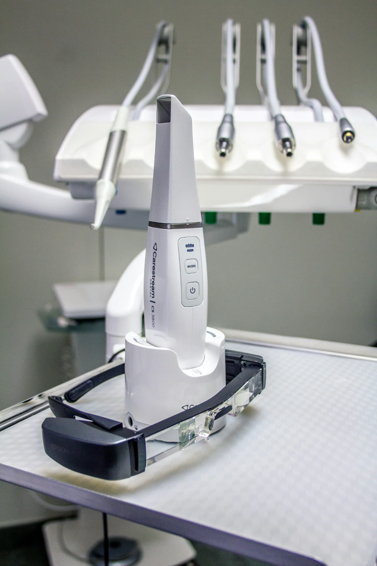
The decision to integrate an intraoral scanner into your practice represents a pivotal moment in your digital journey. It promises enhanced precision, improved patient experiences, and streamlined workflows. But this promise is often clouded by a fog of aggressive marketing, dense technical jargon, and the daunting question of return on investment. How do you separate genuine clinical value from clever sales pitches, especially when considering the high upfront cost and potential for workflow disruption?
This definitive guide is designed to provide clarity. We move beyond manufacturer claims to deliver an expert, unbiased analysis of the technology, its practical clinical integration, and the real-world financial ROI you can expect. We will explore the key differences that matter for specific clinical needs – from restorative to orthodontics, and address the critical concerns of staff training and system interoperability. Our goal is to equip you with the objective insights needed to make a confident investment decision that elevates your practice for years to come.
What is an Intraoral Scanner and How Does It Revolutionize the Dental Practice?
An intraoral scanner is a handheld device that captures direct optical impressions in dentistry. It effectively replaces the conventional tray-and-material method, typically using polyvinyl siloxane (PVS), with a clean, fast, and highly accurate digital workflow. This technology is not merely an alternative to traditional impressions; it represents a fundamental paradigm shift in clinical practice. By creating a precise 3D digital model of a patient's teeth and gingiva, the scanner serves as the essential gateway to the entire ecosystem of digital dentistry, including CAD/CAM milling, 3D printing, and guided implant surgery. The immediate impact is a marked improvement in both clinical efficiency and the patient's perception of the practice as a modern, high-tech provider.
The Shift from Analog to Digital Impressions
The conventional impression process is fraught with challenges. It is often uncomfortable for patients, frequently inducing a gag reflex, and the PVS material itself is susceptible to physical distortions during setting, removal, or shipping. A digital workflow eliminates these variables. The process is reduced from mixing material and waiting for it to set to a simple, non-invasive scan lasting minutes. This transition significantly enhances accuracy, reduces the need for retakes, and eliminates the costs and delays associated with shipping physical models to a dental laboratory.
How the Core Technology Works: An Overview
At its core, an intraoral scanner projects a light source onto the dental anatomy and captures its reflection with imaging sensors. The most common technologies are confocal microscopy and structured light scanning, each designed to rapidly record thousands of individual images. Sophisticated software then "stitches" these images together in real-time to generate a comprehensive 3D surface model. For a more detailed technical Intraoral scanner overview, various academic and reference sources offer in-depth explanations. Early systems required a reflective powder to capture detail, but modern powder-free scanners now dominate the market, further simplifying the clinical process.
Key Benefits for the Practice, Lab, and Patient
The adoption of digital scanning provides distinct, measurable advantages for every stakeholder in the restorative process. The benefits are not isolated but create a cascade of improvements across the entire workflow.
For the dental practice: increased efficiency with reduced chair time per appointment, a significant reduction in material costs, fewer impression remakes, and enhanced diagnostic capabilities through high-resolution 3D models.
For the dental laboratory: instantaneous case reception via secure file transfer, improved and clearer communication with the clinician through digital markups, and higher precision for fabricating restorations.
For the patient: a dramatically more comfortable and faster impression experience, reduced appointment times, and a better understanding of their own oral health through chairside visualization of the 3D scan.
Core Technology: Accuracy, Speed, and Usability
While the market for the modern intraoral scanner is mature, performance disparities remain significant. The underlying technology, a complex interplay of optics, processing power, and software intelligence, directly dictates clinical efficiency, restoration quality, and user satisfaction. Understanding these core technical metrics is essential for differentiating a marketing claim from a tangible clinical advantage and making an informed investment that enhances your digital workflow.
Decoding Accuracy and Precision for Clinical Success
In digital dentistry, accuracy and precision are not interchangeable. Trueness (accuracy) measures how closely the digital model conforms to the patient's actual anatomy, while precision refers to the scanner's ability to produce the same result repeatedly. These factors are non-negotiable, as even micron-level deviations can compromise the marginal fit of crowns, implant restorations, and orthodontic appliances. A scanner's performance here is a function of its optical system, scan strategy, and the power of its software algorithms.
The Importance of Scan Speed and Flow
Scan speed, often measured in frames per second (FPS), translates directly to reduced chair time and improved patient comfort. However, raw speed is only part of the equation. 'Scan flow', the software’s ability to intelligently stitch images and seamlessly resume scanning after interruption, is equally vital. A system that struggles to reorient itself in the presence of saliva or patient movement can quickly negate any on-paper speed advantage, leading to clinical frustration.
Ergonomics and Form Factor: The User Experience
A scanner's physical design has a profound impact on daily usability and operator fatigue. Consider the wand's weight, balance, and button placement, as smaller, lighter designs often improve access to the posterior. System mobility is another key factor; portable laptop-based units offer operatory-to-operatory flexibility, while dedicated cart systems provide a stable, integrated hub. For infection control, practices must choose between systems using autoclavable tips and those reliant on disposable ones.
Finally, the choice between a wired or wireless connection involves a practical trade-off:
Wired scanners: guarantee a stable, uninterrupted data connection and power source, eliminating concerns about battery life and potential signal interference.
Wireless scanners: offer superior ergonomics and freedom of movement, removing cord drag and simplifying handling during full-arch scans.
The optimal choice depends entirely on your practice's specific workflow needs and clinician preference.
Integrating Intraoral Scanners into Daily Workflows
The true value of an intraoral scanner extends far beyond single-unit crown impressions. It serves as the central hub for a fully digital practice, enhancing diagnostic capabilities, improving patient communication, and unlocking new revenue streams. By digitizing the patient's dentition with high-fidelity color and detail, clinicians can visually co-diagnose issues and present treatment plans with greater impact. This digital foundation integrates seamlessly with other equipment like mills and 3D printers, transforming isolated procedures into cohesive, efficient workflows.
Restorative Dentistry: Crowns, Bridges, and Inlays/Onlays
The most immediate application is in chairside CAD/CAM for same-day dentistry. The scan-to-mill workflow allows for the design and fabrication of crowns, inlays, and onlays in a single visit, a significant value proposition for patients. Digital acquisition captures the preparation, opposing arch, and bite registration with exceptional accuracy. Furthermore, a pre-operative scan provides a crucial blueprint for designing anatomically ideal temporary restorations, elevating the standard of provisional care.
Implantology and Surgical Guides
For implantology, the scanner is indispensable. The process involves merging the scanner's STL surface data with a patient's CBCT (DICOM) data. This fused digital model provides the comprehensive information needed for precise virtual implant planning and the subsequent 3D printing of accurate surgical guides. The use of scan bodies ensures the exact three-dimensional position and orientation of the implant are captured, forming the basis for predictable and highly accurate final restorations.
Orthodontics: Clear Aligners and Digital Archiving
In orthodontics, scanners have revolutionized the clear aligner workflow. Digital impressions are submitted directly to providers like Invisalign, eliminating shipping and reducing turnaround times. Advanced software enables powerful outcome simulations, a compelling tool for patient case acceptance. Scanners also facilitate objective progress tracking by overlaying new scans on the initial plan and provide a space-efficient, permanent solution for digital model archiving, replacing cumbersome stone models.
ROI, Pricing Models, and Hidden Costs
The acquisition of a new intraoral scanner represents a significant capital investment, a fact that rightly gives any practice owner pause. However, viewing this technology solely as an expense overlooks its profound potential to drive clinical efficiency and profitability. A rigorous financial analysis reveals that the right scanner is not a cost center, but a strategic investment in the growth and modernization of your practice. The key is to understand the complete financial picture, from initial outlay to long-term value.
Calculating Your Return on Investment (ROI)
A pragmatic ROI calculation moves beyond the initial purchase price to quantify both cost savings and revenue growth. On the savings side, consider the direct elimination of costs for PVS impression materials, trays, and lab shipping. More importantly, the superior accuracy of digital impressions drastically reduces costly and time-consuming remakes. On the revenue side, an efficient intraoral scanner accelerates appointment times, potentially opening up chair time for additional patients. Furthermore, the compelling 3D visuals enhance patient education, leading to higher case acceptance for restorative and cosmetic procedures.
For example, a practice completing 25 crown cases per month could save over $5,000 annually on materials and shipping alone. Factoring in the value of just one additional accepted treatment plan per month transforms the ROI from a long-term goal into a short-term reality.
Purchase vs. Subscription Models
The market offers two primary financial models. A one-time purchase involves a higher upfront capital expenditure but provides ownership and predictable long-term costs, free from recurring fees. Conversely, a subscription model lowers the barrier to entry with a smaller initial payment followed by monthly or annual fees. These subscriptions typically bundle essential services like software updates, technical support, and cloud data access. It is critical to determine which model aligns with your practice's cash flow and long-term financial strategy, as a purchase model may require separate annual fees for software maintenance after the first year.
Ongoing Costs to Consider
The initial quote rarely tells the whole story. Prudent financial planning requires accounting for several ongoing operational costs that are often overlooked:
Consumables: autoclavable scanner tips have a finite number of sterilization cycles before they must be replaced, representing a recurring cost.
Software and support: many manufacturers charge mandatory annual license or support fees after the initial warranty period expires to ensure access to updates and assistance.
Data storage: as cloud-based workflows become standard, be aware of potential monthly fees for data storage and case management platforms.
Training and integration: while initial training is often included, factor in the non-billable time required for your team to adapt and fully integrate the new digital workflow.
To see how these costs stack up for specific models, see our detailed breakdown of the best intraoral scanners and their total cost of ownership.
How to Choose the Right Intraoral Scanner
The preceding reviews offer an in-depth analysis of individual models, but the ultimate question remains: which one is right for your practice? The "best" intraoral scanner is not a universal title; it is the device that most effectively aligns with your clinical needs, workflow philosophy, and long-term business goals. This framework provides a structured, pragmatic approach to making that critical investment.
Open vs. Closed Architecture
This is arguably the most fundamental decision. Open architecture systems allow you to export scans in universal file formats like STL, granting you the freedom to work with any dental lab or design software. In contrast, a closed architecture system locks you into a manufacturer's proprietary ecosystem. While this can offer a seamless, highly integrated workflow for in-office milling, it significantly limits your external collaboration and future flexibility.
Matching the Scanner to Your Clinical Focus
Your primary clinical applications should dictate the features you prioritize. An orthodontic practice requires a scanner with a large field of view for rapid, accurate full-arch scans and excellent color fidelity for patient consultations. For complex restorative or implant work, however, the priority shifts to exceptional margin line sharpness and the ability to capture deep subgingival details with precision. A general practitioner may seek a versatile, all-around performer, whereas a specialist must invest in a tool optimized for their specific procedures.
The Critical Role of Training, Support, and Warranty
Advanced technology is only as valuable as its implementation. Before purchasing, scrutinize the vendor's support infrastructure. Is training conducted on-site by experienced clinicians, or is it a series of online videos? What are the warranty terms, and what is the protocol for a loaner unit if your device requires service? A scanner that is difficult to master or suffers from prolonged downtime becomes a liability, not an asset, regardless of its technical specifications.
To guide your final decision, ask these critical questions:
Does the scanner's architecture (open vs. closed) align with my lab relationship and production goals?
Are its software features and hardware precision suited to my most common clinical procedures?
What is the total cost of ownership, including subscription fees, warranties, and consumables?
How robust is the manufacturer's local support team for training, troubleshooting, and repairs?
Bottom Line
Adopting digital impression technology is no longer a question of 'if,' but 'when and how.' As this guide has detailed, selecting the right intraoral scanner is a practice-altering decision that enhances clinical precision, streamlines restorative workflows, and elevates the patient experience. The key to a successful transition lies in a methodical evaluation that balances core technical performance with the long-term financial and operational impact on your practice.
Frequently Asked Questions
Do intraoral scanners require a powder or coating?
The vast majority of modern intraoral scanners, particularly the leading models reviewed for 2026, are powder-free. This design significantly enhances clinical workflow efficiency and improves patient comfort by eliminating a time-consuming step. While some legacy systems or exceptionally challenging clinical situations involving highly reflective materials might still see marginal benefit from a light dusting agent, for routine restorative and orthodontic cases, powder is now considered an obsolete requirement in digital dentistry.
What is the learning curve for a dentist and their staff to become proficient with an intraoral scanner?
The learning curve has become progressively shorter with advancements in software and hardware. A dentist or assistant can typically achieve basic proficiency for single-unit restorative scans within a few days of hands-on use, supported by initial manufacturer training. Mastering more complex full-arch scans for implant or prosthodontic workflows requires more deliberate practice, often taking several weeks to optimize speed, develop a repeatable technique, and fully integrate the technology into the practice's daily operations.
Can intraoral scanners detect caries or cracks in teeth?
Certain advanced intraoral scanners serve as powerful adjunctive diagnostic tools but do not replace traditional methods like radiographs. Scanners equipped with technologies such as Near-Infrared Imaging (NIRI) can help clinicians visualize potential interproximal carious lesions. Similarly, the high-definition detail of 3D color models can make supra-gingival fracture lines more apparent. However, these features are intended to supplement, not supplant, a comprehensive clinical and radiographic examination for a definitive diagnosis.
How much do the top intraoral scanners cost in 2025/2026?
The investment for a top-tier intraoral scanner system typically ranges from $20,000 to over $45,000. This price often includes the scanning wand and a dedicated laptop or cart. It is crucial for a practice to evaluate the total cost of ownership beyond the initial purchase. Some manufacturers require ongoing annual or monthly subscription fees for software updates, cloud services, and technical support, which must be factored into the overall financial analysis and return on investment.
What is the typical lifespan of an intraoral scanner before it needs to be replaced?
While the physical hardware of a well-maintained scanner can remain functional for over seven years, its clinical lifespan is often dictated by the pace of technological innovation. Most dental practices find themselves upgrading every 5 to 7 years. The primary driver for replacement is typically not mechanical failure but the significant advancements in scan speed, accuracy, color realism, and new software capabilities that make newer models substantially more efficient and clinically valuable for a modern practice.
How do I integrate a scanner with my existing practice management software?
Integration largely depends on the scanner’s architecture. Most contemporary scanners are "open" systems, allowing for the export of standard 3D file formats like STL, PLY, and OBJ. These files can be manually attached to patient records within your practice management software (PMS). For a more seamless workflow, some scanner brands offer direct API integrations with major PMS providers. It is essential to confirm the specific level of compatibility between a prospective scanner and your current software before making a purchase.
Are wireless intraoral scanners as accurate as wired ones?
Yes, for nearly all standard clinical procedures, the accuracy of leading wireless intraoral scanners is clinically equivalent to their wired counterparts. The core data capture technology and optical systems are identical. The decision between wired and wireless is therefore not one of clinical precision but of ergonomics and practice logistics. Wireless models offer superior maneuverability and freedom of movement, balanced against the practical need for battery management and a slightly higher initial investment.