External Cervical Resorption: Diagnosis, CBCT Classification, And Treatment
External cervical resorption (ECR) often hides in plain sight. Early lesions are painless, and the pulp usually tests normal. On a radiograph, ECR can pass for cervical caries or...
Written by Rachel Thompson
Read time: 6 min read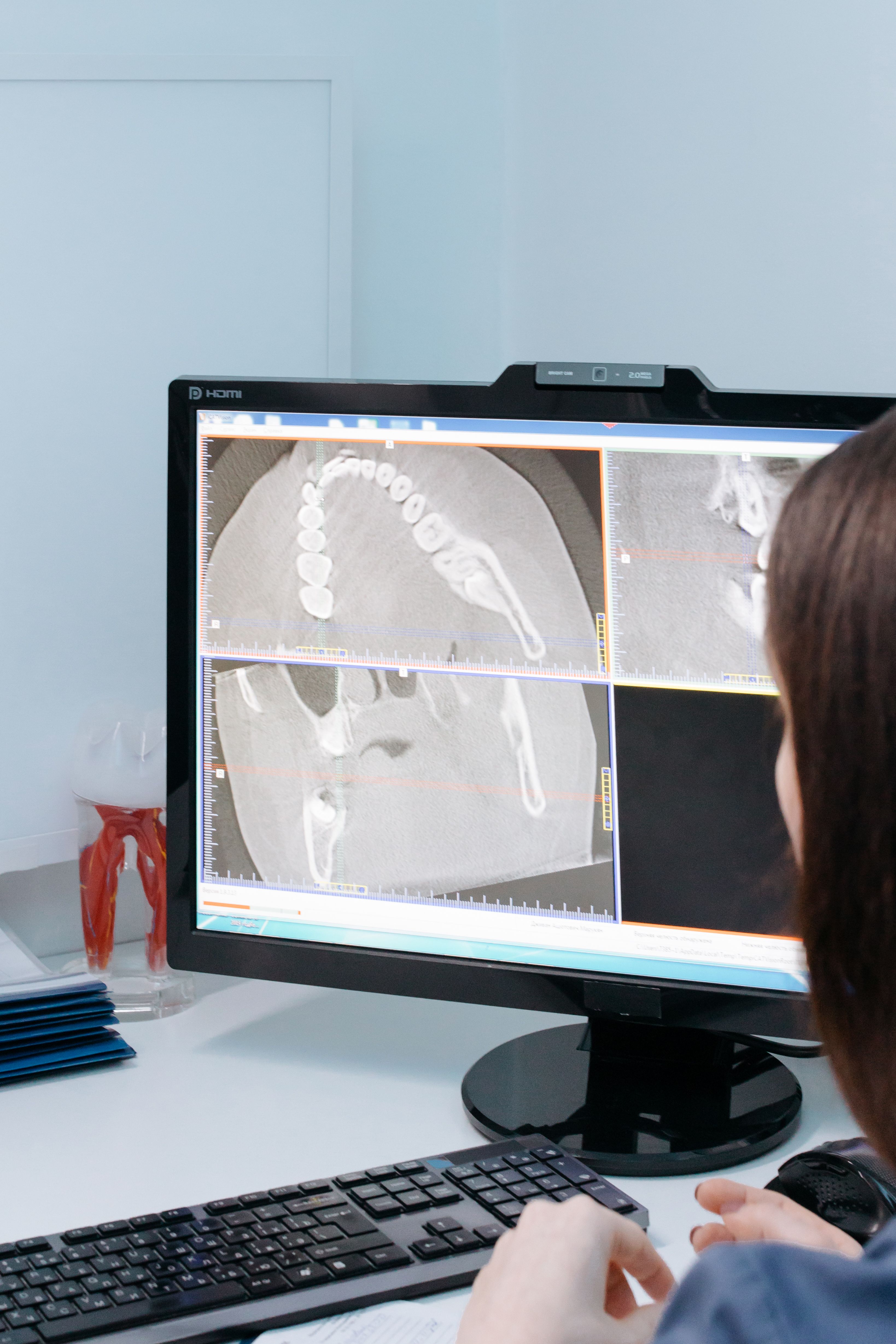
External cervical resorption (ECR) often hides in plain sight. Early lesions are painless, and the pulp usually tests normal. On a radiograph, ECR can pass for cervical caries or internal resorption. Limited-field CBCT and Patel's 3D classification show the true lesion. Its extent decides whether the tooth can be saved.
TL;DR
ECR starts below the gum attachment and spreads through dentin, often without symptoms.
Periapical radiographs underestimate it, so limited-field CBCT guides most treatment plans.
Patel's classification scores height, circumferential spread, and pulp proximity.
Many teeth keep a vital pulp and need repair or monitoring, not root canal treatment.
What Is External Cervical Resorption?
External cervical resorption is a localized, progressive loss of hard tooth tissue. It begins at the cervical root surface, just below the epithelial attachment. From there, it tunnels into dentin and can spread around and along the root.
The condition goes by several names, including invasive cervical resorption and cervical root resorption. The European Society of Endodontology (ESE) position statement uses "external cervical resorption" as the preferred term. ECR is uncommon, but it appears in every age group and on any tooth.
How Does ECR Develop?
Cementum and the cementoid layer normally protect the root surface from resorbing cells. When that layer is damaged, clastic cells reach dentin through a small portal of entry. The lesion then spreads inside the tooth, often far wider than its entry point.
Several features make ECR clinically distinctive:
A small portal of entry with a much larger lesion inside
Irregular channels that interconnect through the dentin
A thin layer of dentin and predentin that resists resorption around the canal
Bone-like reparative tissue laid down in older lesions
That protective layer around the canal is called the pericanal resorption-resistant sheet. It explains why many large lesions still have a vital, normally responding pulp. Reparative tissue explains why some lesions look partly radiopaque and seem to slow down.
ECR Vs. Other Types Of Root Resorption
ECR is one of several resorption types. The 2023 ESE position statement on root resorption separates them by origin and behavior.
Type | Where it starts | Typical driver |
|---|---|---|
External cervical resorption | Cervical root, below the attachment | Damaged root surface, often no infection |
External inflammatory resorption | Any root surface, often after trauma | Infected, necrotic pulp |
Replacement resorption | Root surface fused to bone | Ankylosis after severe trauma |
Internal resorption | Inside the canal wall | Inflamed but partly vital pulp |
The practical difference lies in the pulp. External inflammatory resorption stops when the infected canal is treated. ECR does not depend on the pulp, so root canal treatment alone will not stop it.
What Causes External Cervical Resorption?
The exact cause of ECR is still unknown. Researchers describe predisposing factors, which are associations rather than proven causes. Many patients have more than one, and some have none.
Common Predisposing Factors
A 2025 observational study of 215 teeth by Patel and colleagues recorded these factors at diagnosis:
Orthodontic treatment, in 25.6% of teeth
Dental trauma, in 20.9% of teeth
Domestic cat ownership, in 15.8% of teeth
Parafunctional habits, in 10.2% of teeth
No identifiable factor, in 22.3% of teeth
Internal bleaching, periodontal surgery, and other injury near the cementoenamel junction also appear in case histories. The cat-ownership link is the most unusual finding. A feline virus has been proposed as a trigger, but no causal link has been shown.
What This Means For History-Taking
A focused history helps when a cervical defect looks unusual. Clinicians can ask about orthodontics, past trauma, bleaching of a dark tooth, and periodontal surgery. Pets and grinding habits now belong on that list too.
The history also guides screening. Patients with multiple factors deserve a closer look at cervical areas on routine bitewings. Multiple lesions in one mouth raise the possibility of multiple idiopathic cervical resorption, a rarer pattern.
What Are The Signs And Symptoms Of External Cervical Resorption?
Most early ECR is found by chance on routine radiographs. The tooth usually feels normal. Pain rarely appears until the pulp or periodontium is involved.
Clinical Findings To Look For
When signs appear, they are often subtle:
A cervical cavity with hard, sharp margins
Heavy bleeding when the defect is probed
A gritty, scratchy feel under the probe
A localized pocket that does not match the rest of the mouth
Pink discoloration of the crown in some coronal lesions
The classic "pink spot" appears only when vascular tissue lies close under thin enamel. Most ECR lesions never show it. A defect that bleeds briskly yet feels hard and sharp-edged should raise suspicion. Cervical caries feels soft and sticky by comparison.
Why Pulp Tests Often Stay Normal
The resorption-resistant sheet shields the pulp until late in the disease. Cold and electric tests often give normal responses, even with large lesions.
A normal pulp test therefore does not rule out ECR. Symptoms of pulpitis or apical periodontitis usually mean the lesion has finally breached the canal. Those cases change the treatment plan and the pulp prognosis.
How Is External Cervical Resorption Diagnosed?
Diagnosis combines clinical examination, conventional radiographs, and, where it changes management, limited-field CBCT. Each step answers a different question.
Clinical Examination And Pulp Testing
The exam starts with careful circumferential probing, ideally under magnification. Clinicians should map the defect, note bleeding, and check existing restorations at the gingival margin. Pulp sensibility tests and percussion establish a baseline, and the history covers known predisposing factors.
Probing has limits. Interproximal and palatal portals of entry are easy to miss, especially under contact points. A lesion felt as a small catch can turn out to be large on imaging.
Periapical Radiographs And Their Limits
On a periapical radiograph, ECR usually looks like a mottled, poorly defined cervical radiolucency. Two radiographic signs help separate it from internal resorption:
The canal outline stays visible through the ECR lesion
The lesion shifts position on a second, angled view
Internal resorption balloons out from the canal and stays centered on it in both views. This parallax technique is quick and should come before any CBCT. Two-dimensional images still miss depth and circumferential spread. Our guide to dental radiographic imaging covers the basics of these views.
When Is CBCT Recommended For ECR?
Limited-field CBCT shows the lesion in three planes. The 2023 ESE position statement supports it for localizing resorptive defects and planning treatment. It reveals details a periapical image cannot:
The location of the portal of entry
How far the lesion spreads around the root
Its height relative to the bone crest
Its distance from the root canal
Areas of reparative, bone-like tissue
CBCT comes after a clinical exam and periapical views. It is justified when the answer could change treatment. A small, supracrestal buccal lesion seen clearly on probing may not need a scan. Our CBCT scanning in dentistry guide explains the technology and dose. Our CS 9600 review covers one limited-field option.
External Cervical Resorption Classification: Heithersay Vs. Patel
Two systems classify ECR. They measure different things and should not be treated as interchangeable.
Heithersay Classification: Classes 1–4
Heithersay's system, introduced in 1999, grades lesions on two-dimensional radiographs. It describes how deep the lesion has spread into the tooth:
Class 1: Small cervical lesion with shallow dentin penetration
Class 2: Well-defined lesion close to the pulp chamber, with little radicular spread
Class 3: Deeper invasion into the coronal third of the root
Class 4: Large lesion extending beyond the coronal third of the root
A 2026 narrative review pooled 657 teeth graded with Heithersay's system. Classes 1 and 2 had high survival, while Class 4 had poor outcomes. The system is simple, but it cannot show how far a lesion wraps around the root.
Patel's Three-Dimensional CBCT Classification
Patel and colleagues' classification uses CBCT to score three features. Each lesion gets a number, a capital letter, and a lowercase letter.
Component | Score | Meaning |
|---|---|---|
Height | 1 | At the cementoenamel junction or supracrestal |
Height | 2 | Coronal root third, below the bone crest |
Height | 3 | Extends into the middle root third |
Height | 4 | Extends into the apical root third |
Circumferential spread | A | Up to 90° |
Circumferential spread | B | More than 90° up to 180° |
Circumferential spread | C | More than 180° up to 270° |
Circumferential spread | D | More than 270° |
Root canal proximity | d | Lesion confined to dentin |
Root canal proximity | p | Probable pulpal involvement |
Reading a classification: 2Bp
A 2Bp lesion sits in the coronal root third below the crest. It spreads more than 90° but no more than 180° around the root. Pulp involvement is probable. A 2026 case report described a 2Bp incisor repaired from inside the canal. It remained stable after a decade.
Why Three-Dimensional Classification Matters
A 3D code gives the whole team the same picture of the lesion. It supports several practical tasks:
Clear referral letters between general dentists and endodontists
Planning surgical or internal access to the lesion
Estimating how much sound tooth structure will remain
Tracking lesion progression on follow-up scans
The evidence on prognosis is still forming. A 2026 multi-center study found that Patel scores tracked survival more closely than Heithersay classes. Spread beyond 180° carried the highest extraction risk. The 2026 narrative review noted that studies using both systems were small with short recalls. A Patel code describes a lesion and does not guarantee an outcome.
How Is External Cervical Resorption Treated?
Treatment aims to stop the resorptive process, seal the defect, and keep the tooth functional. The right approach depends on access, lesion extent, pulp status, and periodontal support. Recording the reasoning in a structured dental treatment plan helps when cases are reviewed years later.
External Surgical Repair
External repair suits lesions the surgeon can reach from the root surface. Buccal and many palatal lesions in the coronal third fall into this group. The usual sequence runs as follows:
Raise a flap or retract the gingiva to expose the portal of entry.
Remove all resorptive tissue with burs, curettes, and magnification.
Apply 90% trichloroacetic acid in selected cases to reach tissue in deep channels.
Treat the pulp if the canal is exposed or infected.
Restore the defect and reposition the soft tissue.
Subcrestal lesions are harder. Moisture control becomes difficult, and the restoration margin ends up below the bone. Orthodontic extrusion or crown lengthening can bring the margin into a cleansable position. In the esthetic zone, that trade-off needs a frank discussion with the patient before surgery.
Internal Repair
Internal repair reaches the lesion through an access cavity in the crown. It suits lesions that cannot be reached surgically, such as interproximal or deep subcrestal defects. The clinician removes resorptive tissue from inside, then seals the defect, often with a calcium silicate material.
This approach differs from routine obturation. Its goal is sealing a perforating defect inside the root. A 2026 Journal of Endodontics case report described eight ECR teeth managed partly through the canal. It kept the option of later surgical repair open. Our root canal anatomy article covers the terminology used in these cases.
When Is Root Canal Treatment Necessary?
An ECR diagnosis alone does not call for root canal treatment. Endodontic treatment is indicated when:
The pulp is necrotic or irreversibly inflamed
Removing the lesion exposes the canal
Internal repair requires canal access
Apical periodontitis is present
Teeth with a "d" proximity score and normal pulp tests are good candidates for pulp preservation. Even small exposures during repair can sometimes be managed with vital pulp therapy. Our guide to minimally invasive dentistry covers pulp-preserving approaches in more depth.
When Are Monitoring Or Extraction Considered?
Monitoring is a deliberate choice for selected lesions. It may suit these situations:
Lesions with extensive reparative, bone-like tissue
Lesions where access would cost more tooth than the disease has
Patients who decline surgery after informed discussion
Medically complex patients for whom surgery carries high risk
A 2026 case report described an ECR lesion that filled in with reparative tissue without treatment. The authors listed four possible outcomes of monitoring, from stability to progression requiring extraction. Extraction becomes the realistic option when the spread is extensive, and the remaining root cannot support a restoration. Implants or other replacements then enter the plan.
What Materials Are Used To Repair External Cervical Resorption?
No single material suits every ECR defect. The choice depends on margin position, moisture control, and contact with pulp or periodontal tissue.
Matching The Material To The Defect
Each material group has a typical role in ECR repair:
Resin composite: supragingival margins with good isolation and esthetic demands
Glass ionomer or resin-modified glass ionomer: subgingival margins in contact with soft tissue
Calcium silicate cements: pulp-near areas, perforations, and internal repairs
Many clinicians layer materials. Glass ionomer sits against the gingiva and composite covers the visible surface. Calcium silicate materials such as Biodentine tolerate moisture and support pulp healing. Our Biodentine in dentistry guide and Biodentine review cover the material in detail.
Restoring Strength After Repair
A large cervical defect weakens the tooth at its most loaded point. A 2025 Journal of Endodontics study tested 81 incisors with simulated ECR defects connected to the pulp. Restorative material choice changed fracture resistance, while buccal or palatal location did not.
Root fracture is a common late failure. Clinicians should plan occlusal protection and full coverage when the defect undermines the crown. Posts are best avoided where they could interfere with a calcium silicate repair.
Clinical Decision Framework For External Cervical Resorption
The table links common findings to the questions they raise. It is an educational framework and does not replace an individual endodontic assessment.
Clinical finding | Treatment-planning consideration |
|---|---|
Small, accessible cervical lesion | Assess feasibility of external repair |
Extensive circumferential spread | Evaluate remaining tooth structure and restorability |
Lesion approaching the pulp | Use clinical findings and CBCT to assess pulp involvement |
Probable or confirmed canal communication | Consider whether endodontic treatment is indicated |
Subcrestal extension | Evaluate periodontal access and restorative sealing |
Extensive structural destruction | Assess prognosis, extraction, and replacement options |
Uncertain lesion activity | Plan clinical and radiographic follow-up |
Questions To Answer Before Choosing Treatment
Five questions settle most ECR plans. Working through them in order keeps the decision honest.
Can the whole lesion be reached and cleaned?
Will enough sound tooth remain to restore and protect?
Is the pulp healthy, inflamed, or infected?
Can the restoration margin be kept clean and periodontally stable?
Does the patient accept the effort, cost, and long-term uncertainty?
A "no" to the first two questions usually points toward monitoring or extraction. Complex cases benefit from early referral to an endodontist with CBCT and a microscope.
What Is The Prognosis For Teeth With External Cervical Resorption?
Prognosis depends on lesion extent, remaining tooth structure, pulp status, and periodontal support. Recent studies report useful numbers, but no single survival rate applies to every case.
What Do Recent Outcome Studies Show?
Studies differ in design, case mix, and follow-up, so their figures should not be pooled.
Study | Design | Key outcome |
|---|---|---|
Patel et al., 2026 | 296 teeth, 5 specialist centers | 85.7% survival at 5 years, 80.5% at 10 |
Pannkuk, 2026 | Single operator, 1996–2023 | 89.5% 5-year survival in Heithersay Class 2 |
Mota de Almeida et al., 2026 | 14,881 teeth, Swedish registry | 82.7% 5-year, 74.9% 10-year survival after conservative care |
Fernández-Grisales et al., 2026 | External surgical repair only | 77.8% survival at median 5.6 years |
What Affects The Outcome?
Several patterns repeat across these studies:
Greater lesion extent predicts lower survival
Spread beyond 180° carried the highest risk in the multi-center study
Anterior teeth had lower 5-year survival than molars in the single-operator series
Root fracture was a leading reason for late extraction
Treatment comparisons need care. In the multi-center study, internal repair had the lowest estimated survival at 59.1%. Clinicians chose internal repair for less accessible lesions. The figure reflects case selection as much as technique. The Swedish registry also found 42.8% of ECR teeth were extracted at first treatment.
Follow-up should extend for years. The Colombian cohort found critical time points at 2 and 3 years after repair. Success judged on CBCT was 49.3%, versus 61.7% on periapicals, a non-significant difference. Selected cases may warrant a follow-up scan when periapicals look stable, but symptoms persist.
Bottom Line
External cervical resorption is easy to miss. Early lesions are silent, and periapicals hide their true size. A careful probe, a parallax view, and limited-field CBCT reveal what the tooth is facing. Patel's 3D classification turns that image into a shared plan. Repair, pulp treatment, monitoring, or extraction still depends on access, remaining structure, and patient goals.
Frequently Asked Questions
What is external cervical resorption?
It is a progressive loss of hard tooth tissue starting just below the gum attachment. It spreads into the dentin and around the root. Our CBCT scanning in dentistry guide explains how 3D imaging shows it.
Can external cervical resorption be reversed?
Lost tooth structure does not grow back. Treatment removes the resorptive tissue and seals the defect. Some lesions also lay down reparative tissue on their own. Materials such as those in our Biodentine guide help seal the repair.
Does external cervical resorption always require a root canal?
No. Many teeth keep a healthy pulp and need only repair or monitoring. Root canal treatment is needed when the pulp is infected, necrotic, or exposed during repair. Our root canal anatomy article covers related terms.
Can CBCT detect external cervical resorption?
Yes. Limited-field CBCT shows the lesion's height, spread around the root, and distance from the canal. It works best after a clinical exam and periapical radiographs.
Can a tooth with external cervical resorption be saved?
Often, yes, especially when the lesion is found early and is accessible. Extensive spread and weak remaining structure lower the odds. A documented dental treatment plan helps patients weigh repair against extraction.