Mucocele In Mouth: Causes, Symptoms, And Treatment
A soft, painless bubble inside the lower lip often shows up after an accidental bite. It is often a mucocele, a pool of saliva leaking from a damaged minor salivary gland. Most...
Written by Agnes Markovic
Read time: 6 min read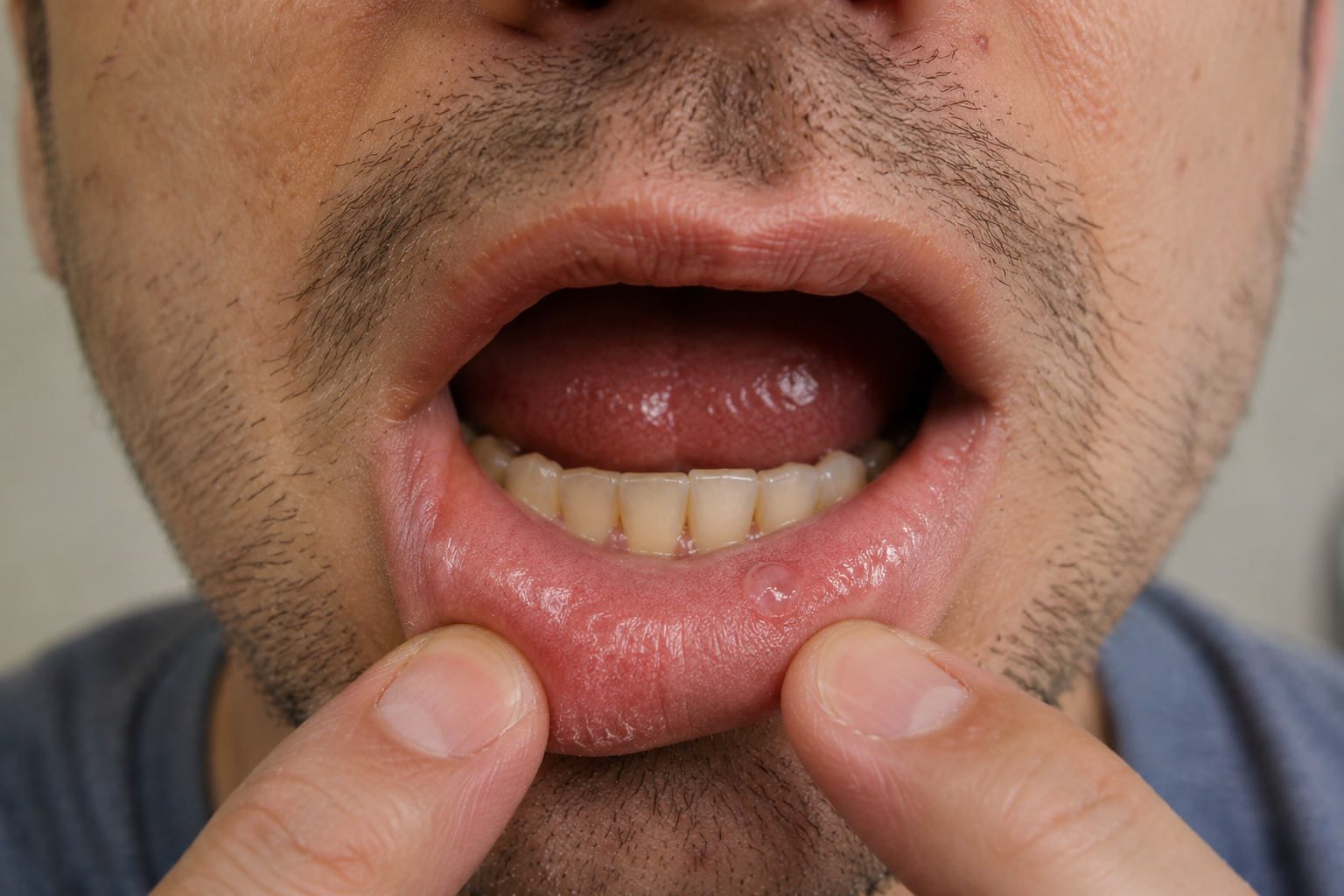
A soft, painless bubble inside the lower lip often shows up after an accidental bite. It is often a mucocele, a pool of saliva leaking from a damaged minor salivary gland. Most heal without treatment. A lump that lingers, grows, or keeps coming back needs a dental exam.
TL;DR
A mucocele forms when an injured minor salivary gland leaks saliva into nearby tissue.
Accidental lip biting causes most cases, so the inner lower lip is the usual spot.
Many mucoceles burst and heal on their own within 3 to 6 weeks.
Book a dental exam if the bump lasts past two weeks, keeps returning, or changes in feel or color.
What Is An Oral Mucocele?
An oral mucocele is a soft swelling filled with thick saliva. It comes from one of the minor salivary glands. These tiny glands sit under the lining of the lips, cheeks, tongue, and palate. Each drains through a short duct.
A mucocele forms in one of two ways. Extravasation mucoceles appear when saliva leaks out of a torn duct. The body walls off the pool with granulation tissue, so no true lining forms. Pathologists call this a pseudocyst. Retention mucoceles appear when a blocked duct swells with trapped saliva. The duct lining surrounds the fluid, which makes it a true cyst.
Mucoceles are benign and do not spread from person to person. A review of 1,824 cases found an average patient age of about 25 years. Males and females were affected about equally.
What Does A Mucocele In The Mouth Look And Feel Like?
A typical mucocele looks like a small, smooth blister just under the surface. Most measure a few millimeters to about a centimeter, though some reach 2 centimeters. Common features include:
Round or dome-shaped swelling
Soft, rubbery, or fluid feel when pressed
Clear, bluish, pink, or the same color as nearby tissue
Usually painless, though repeated biting can make it sore
A cycle of bursting, draining clear, sticky fluid, and refilling
Color depends on depth. Shallow lesions let light pass through the fluid, which creates the bluish tint. Deeper ones look like a normal pink bump. In a study of 105 mucoceles, 63 had normal coloring, and only 34 looked bluish. A pink bump can still be a mucocele, and a bluish bump can be something else. Many patients notice it swells around meals, when saliva flow rises. It then settles a few hours later.
Mucocele Inside The Lower Lip
The inner lower lip is by far the most common site. In a study of 89 children, 79.8 percent of mucoceles sat on the lower lip. This lip rests in the path of the lower front teeth during chewing. A new mucocele often follows a recent change. New braces, a chipped front tooth, or a new crown are common triggers. Braces add more risk, and orthodontic wax over sharp brackets cuts down on rubbing.
The upper lip rarely develops mucoceles. A swelling there is more likely to be a different lesion. Dentists check upper lip lumps closely for salivary gland tumors.
Mucocele Under The Tongue Or Elsewhere In The Mouth
Mucoceles also form on the inner cheeks, the palate, and the underside of the tongue tip. A ranula is a related swelling on the floor of the mouth. It usually comes from the sublingual gland, a major salivary gland. A plunging ranula can push through the floor-of-mouth muscle into the neck. Ranulas often need imaging and a different surgical plan.
What Causes A Mucocele In The Mouth?
Every mucocele starts with a duct that stops draining properly. Many people recall no injury at all. In the 105-case study, 51 patients reported no trauma or biting habit. Small bites during meals often go unnoticed.
Accidental Lip Biting And Oral Trauma
A hard bite while chewing can tear a duct in an instant. The bump may take a few days to appear. Falls, sports injuries, and dental procedures can damage ducts the same way.
Blocked Salivary Gland Ducts
Retention mucoceles form when something plugs the duct. Thick mucus, old scar tissue, or a tiny salivary stone can block the exit. The gland keeps making saliva, so the duct balloons. In a study of 58 cases, only 15.5 percent were retention mucoceles.
Repeated Irritation And Oral Habits
Some people chew or suck their lower lip when stressed or focused. That pressure reinjures the same glands, so mucoceles return to the same spot. A lip-biting or oral fixation habit is worth addressing. Sharp tooth edges and rough fillings can also rub the lip all day. Poor brushing and bacteria do not cause mucoceles.
Types Of Oral Mucoceles
Oral mucoceles fall into two main types based on how the saliva escapes. A lab confirms the type after removal.
Type | How it forms | Main distinction |
|---|---|---|
Extravasation mucocele | Saliva leaks out of a torn duct into connective tissue | Far more common, often linked to trauma, no true lining |
Retention mucocele | Saliva builds up behind a blocked duct | True cyst lined by duct cells, seen more in older adults |
A third variant, the superficial mucocele, looks like a tiny, tense, clear blister. It favors the soft palate, the back of the cheeks, and the area behind the molars. It often bursts within a day or two. A 2023 review notes these were long misdiagnosed as blistering diseases, such as mucous membrane pemphigoid. Clusters of blisters with redness and burning may point toward stomatitis instead.
How Is An Oral Mucocele Diagnosed?
Dentists diagnose most mucoceles during a regular exam. The look, feel, and history usually settle it. The dentist still rules out other masses. During the exam, a dentist checks:
Location, size, color, and texture
Recent lip bites, falls, dental work, or chewing habits
How long the bump has been present
Whether it has burst and refilled
Gentle pressure tells a lot. A soft swelling with a fluid feel fits a mucocele. A firm lump that holds its shape raises other possibilities. A blood vessel lesion may blanch under pressure, and a mucocele does not. Dentists record size and color in the dental treatment plan. That record gives the recheck a clear baseline.
When Is A Biopsy Necessary?
Most removed mucoceles go to a lab to confirm the diagnosis. A biopsy before treatment makes sense when the lesion breaks the classic pattern:
It feels firm or fixed to deeper tissue
It grows steadily or changes color
The surface becomes a sore that will not heal
It sits on the upper lip or another unusual site
Imaging is rarely needed for a small lower lip lesion. Ultrasound, CT, or MRI is mostly reserved for ranulas and deep swellings.
Mucocele Vs. Canker Sore, Fibroma, And Other Mouth Bumps
Several mouth lesions look alike in the first few days. Texture and pain are the most useful early clues. The table lists typical patterns only.
Condition | Typical distinguishing feature |
|---|---|
Oral mucocele | Soft, smooth, fluid-filled bump, often on the inner lower lip |
Canker sore | Painful, shallow ulcer with a pale center and red border |
Irritation fibroma | Firm, solid growth from long-term rubbing or biting |
Dental abscess | Pain, pus, or a gum boil near an affected tooth |
Other salivary gland lesions | Can mimic a mucocele and may need specialist review |
A canker sore is an open, painful ulcer. A mucocele is an intact, fluid-filled bump. The guide to canker sores and mouth ulcers covers those sores. A gum boil near an aching tooth points to a tooth abscess. Flat white patches are associated with white spots on gums. Appearance alone cannot rule out a tumor.
Do Oral Mucoceles Go Away On Their Own?
Yes, many do. The swelling bursts, the fluid drains, and the duct heals. Most run their course in 3 to 6 weeks. In the study of 89 children, 43.8 percent cleared without treatment.
Bursting does not always mean healing. If the duct stays damaged, the bump refills. Each cycle can leave scar tissue that makes the lump firmer.
Do not pop, pierce, or cut a mucocele at home. A pin or blade can introduce bacteria and injure nearby glands. That damage can make the problem return.
Oral Mucocele Treatment: When Is Removal Necessary?
Treatment depends on size, location, duration, and recurrence. Dentists usually treat a mucocele that persists or interferes with eating and speaking.
Observation And Conservative Management
A small, painless mucocele after a known bite often gets a watch-and-wait plan. The dentist measures it and sets a recheck date. Avoid biting or poking the area. Report any growth, firmness, or color change sooner.
Surgical Mucocele Removal
Surgical excision is the standard approach for persistent lesions. The dentist or oral surgeon numbs the area. They remove the swelling along with the minor salivary glands feeding it. Recurrence drops sharply once that gland tissue is fully removed. Draining the fluid with a needle alone usually fails because the injured duct keeps leaking. The tissue then goes to a pathology lab.
Laser Treatment And Other Procedures
CO2 and diode lasers can remove the lesion with less bleeding. Cryotherapy freezes the tissue. Micro-marsupialization passes a suture through the dome to create new drainage paths. It suits young children who may not tolerate surgery. A 2023 meta-analysis found no clear difference in recurrence between scalpel, CO2 laser, and marsupialization. The choice depends on the lesion, patient age, and clinician training.
What Happens After Mucocele Removal?
Recovery after a small excision is usually short and mild. Healing time varies with lesion size and technique. Typical aftercare includes:
Mild tenderness and swelling for a few days
Soft foods and lukewarm drinks while the site heals
Gentle brushing that avoids the stitches
A follow-up visit to check healing and recurrence
Many clinicians suggest a saltwater mouth rinse after the first day. For soreness, read our comparison on whether Tylenol or Ibuprofen is better. Fine sensory nerve branches run just under the lip glands. A small patch of numbness can occur and usually fades over a few weeks.
Can A Mucocele Come Back After Treatment?
Yes, though most do not after complete removal. Recurrence usually traces back to leftover gland tissue or ongoing lip trauma.
Reported rates vary by study and technique. A study of 68 patients found recurrence in 8.8 percent of scalpel removals. In the pediatric study, 8 percent of removed lesions came back. Small samples like these do not give a universal rate. A new bump near the scar needs a recheck.
When Should You See A Dentist For A Mouth Mucocele?
Use the framework below as a timing guide, not a self-diagnosis tool. Any doubt about a mouth lump is reason enough to book an exam.
Reasonable To Watch Briefly
Short observation fits a bump that matches the classic pattern. All of these should apply:
It appeared soon after a known lip bite
It is soft, smooth, and painless
It sits on the inner lower lip
It is shrinking or has burst and flattened
Book A Dental Exam
Schedule a visit if the lump shows any of these signs. Do not wait out the full 3 to 6 weeks.
It lasts more than about two weeks without clear improvement
It returns or grows larger
It feels firm, bleeds, ulcerates, or changes color
It hurts or makes speaking, chewing, or swallowing harder
The ADA's 2026 living guideline treats reassessment of persistent lesions as part of diagnosis. It calls a recheck 10 to 14 days after finding a suspicious lesion medically necessary. The new oral cancer screening guidance covers this in detail.
Get Urgent Care
Some swellings need same-day medical attention. Go to urgent care or an emergency room for:
Fast-growing swelling under the tongue or on the floor of the mouth
Trouble breathing or swallowing
Swelling that spreads into the neck or jaw
Bottom Line
An oral mucocele is a harmless pocket of saliva from an injured or blocked gland. Most appear on the inner lower lip after a bite. Many burst and heal within weeks. Persistent, recurring, painful, or unusual lumps need a dental exam. Removing the lesion and its feeding glands resolves most stubborn cases. A bluish color alone does not confirm a mucocele, so let a dentist make the call.
Frequently Asked Questions
Is an oral mucocele dangerous?
No, a true mucocele is benign. The risk lies in mistaking another lesion for one. Any lump that lasts past two weeks or feels firm needs an exam. Routine oral cancer screening catches lesions that only look harmless.
How long does a mucocele last?
Many last 3 to 6 weeks before they burst and heal. Some clear within days. Others persist for months or keep returning because the duct stays damaged. A bump that keeps refilling usually needs removal of the feeding gland tissue.
Can I pop a mucocele in my mouth?
No. Popping or cutting it at home can cause infection and injure nearby glands. Draining it also leaves the leaking duct unrepaired, so it usually refills. A dentist can remove it in a short visit under local anesthesia.
Is a mucocele contagious?
No, a mucocele is not an infection. It forms from saliva leaking out of an injured gland. It cannot pass through kissing or shared utensils. Contagious sores look different, as the guide to canker sores, cold sores, and mouth ulcers explains.
Can a mucocele turn into cancer?
No, a true mucocele does not become cancer. Some tumors can look similar at first, though. Dentists send removed tissue to a lab for that reason. A lump with an unclear history may need a biopsy to confirm what it is.
Why does my mucocele keep coming back?
Repeated injury to the same gland is the usual cause. Lip biting, sharp teeth, or braces can keep damaging the duct. Incomplete removal of the feeding gland is another reason. Wax over sharp brackets and help with an oral fixation habit both lower the odds.
Do I need surgery for an oral mucocele?
Not always. Small mucoceles that shrink after an injury often heal without treatment. A procedure makes sense when the lump persists, recurs, or interferes with eating. If removal is advised, learn how to read a dental treatment plan before agreeing.