ADA Releases New Oral Cancer Screening Guidelines
The 2026 ADA oral cancer guidelines keep the clinical exam at the center of early detection. The ADA published its final recommendations in JADA on September 28. Suspicious or...
Written by Rachel Thompson
Read time: 4 min read
The 2026 ADA oral cancer guidelines keep the clinical exam at the center of early detection. The ADA published its final recommendations in JADA on September 28. Suspicious or persistent lesions need prompt biopsy or referral. Screening adjuncts should not decide that step. This article explains what the guideline means at the chair.
TL;DR
The ADA recommends a full extraoral and intraoral exam for every adult at every visit type
Dentists should biopsy or refer suspicious lesions promptly, plus any lesion unresolved after 10 to 14 days
The guideline advises against routine cytology, vital staining, light-based, and salivary adjuncts for biopsy decisions
Practices need a tracked follow-up system so no unresolved lesion waits for the next recall
What Changed In The 2026 ADA Oral Cancer Guidelines?
The 2026 ADA oral cancer guidelines recommend a comprehensive extraoral and intraoral exam for all adults. Suspicious lesions need a prompt biopsy or specialist referral. A punch or scalpel biopsy with histopathology remains the definitive diagnostic method. It advises against routine cytology, vital staining, light-based, and salivary adjuncts for biopsy decisions.
The ADA oral cancer guideline updates the ADA's 2017 guideline on evaluating potentially malignant disorders. It covers oral and lip squamous cell carcinoma (OSCC) and oral potentially malignant disorders (OPMDs). The ADA released it in four parts during 2026. The final set of recommendations appeared online in JADA on September 28.
It is also the first guideline completed under the ADA Living Guideline Program, which launched in 2025. Traditional guidelines typically update every three to five years. A living guideline updates as soon as new evidence emerges. Methodologists from Penn Dental Medicine's Center for Integrative Global Oral Health helped develop it. The ADA Council on Scientific Affairs approved the final text.
Which Adjuncts Did The Guideline Evaluate?
The panel reviewed four categories of chairside adjuncts. Each one was judged on whether it should guide the decision to biopsy or refer.
Cytology, which samples loose surface cells from a lesion
Vital staining, such as toluidine blue, which dyes suspicious tissue
Light-based adjuncts, including autofluorescence and tissue reflectance devices
Salivary adjuncts, which test saliva for disease-related biomarkers
What Stayed The Same?
Most of the clinical core carries over from earlier ADA guidance. The exam and the biopsy remain the two fixed points of early detection.
The update adds current evidence on each adjunct and a clear time trigger for unresolved lesions. It also frames follow-up visits as part of the diagnostic process. A dentist who already examines every adult and biopsies promptly will change little at the chair. The bigger changes sit in tracking, documentation, and adjunct use.
Clinical Examination Remains The Foundation Of Oral Cancer Detection
The guideline asks for a comprehensive extraoral and intraoral exam on every adult patient. ADA News reports that this applies at new-patient, recall, and emergency visits alike.
The emergency visit deserves attention. A patient in pain often skips recalls, and the urgent appointment may be the only exam that year. Irregular attenders who also use tobacco or alcohol heavily carry a higher oral cancer risk.
What Does A Complete Oral Cancer Examination Include?
A complete exam combines sight and touch across the head, neck, and mouth. Palpation finds induration and fixed masses that look normal on the surface.
The extraoral part checks facial symmetry, skin, lip vermilion, and cervical and submandibular nodes. The intraoral part covers every mucosal surface in a fixed order:
Labial and buccal mucosa
Gingiva and alveolar ridges
Dorsal, lateral, and ventral tongue
Floor of the mouth
Hard and soft palate
Oropharynx and tonsillar pillars
The lateral and ventral tongue and the floor of the mouth need a dry field and good retraction. Gauze on the tongue tip helps pull it to each side. A fixed sequence in the dental charting workflow keeps sites from being skipped.
Which Histories Need Updating?
The guideline asks clinicians to update medical, social, and dental histories at each exam. Each history adds context to what the exam finds.
The social history records tobacco, alcohol, and betel quid use. Smoking and vaping histories should note product type and duration. Sun exposure matters for lip lesions, and HPV status matters for oropharyngeal findings. The medical history flags immunosuppression and any prior head and neck cancer.
What Findings Should Raise Concern?
Some findings need closer evaluation regardless of the patient's risk profile. These include:
White or red patches that do not wipe off
Mixed red and white lesions
Ulcers with no clear cause, or ulcers that persist
Induration, fixation, or a mass on palpation
Unexplained numbness, bleeding, or loose teeth
Persistent hoarseness, sore throat, or trouble swallowing
Many lesions have a benign explanation, such as trauma or a canker sore. A plausible cause still needs confirmation through healing. That is where the guideline's 10- to 14-day window applies.
When Should A Dentist Biopsy Or Refer An Oral Lesion?
The guideline gives four clear triggers for moving a lesion toward definitive diagnosis. Each one shortens the time between first sighting and a pathology report.
Suspicious Lesions
A lesion that looks or feels suspicious needs a biopsy without delay. The guideline says clinicians should perform it promptly or refer the patient to a specialist.
Referral options include an oral and maxillofacial surgeon, an oral medicine specialist, or an oral pathologist. The referral letter should state the site, size, duration, and why the lesion raised concern.
Lesions Without A Definitive Diagnosis
Many lesions have a likely benign cause, such as a sharp cusp or denture flange. The guideline sets a firm checkpoint for these cases.
A lesion unresolved 10 to 14 days after the initial evaluation needs prompt biopsy or referral. This applies when there is still no definitive diagnosis or treatment plan. The clock starts at the dentist's evaluation. Patients often report a lesion only after it has been present for some time. The ADA's patient advice flags any sore or lump unhealed after two to three weeks.
Lesions With A Negative Adjunct Result
A negative adjunct result does not close the case. The guideline addresses this directly for cytology.
A lesion that persists or progresses after negative cytology needs immediate biopsy or referral. A clinically worrying lesion outranks a reassuring test.
Punch Or Scalpel Biopsy For Definitive Diagnosis
Only tissue confirms the diagnosis. Punch or scalpel biopsy with histopathology is the preferred definitive method for OSCC and OPMDs.
Incisional biopsy of the most suspicious area suits larger or mixed lesions. Excisional biopsy suits small, well-defined lesions. Dentists who do not perform biopsies should refer early, before the review window closes.
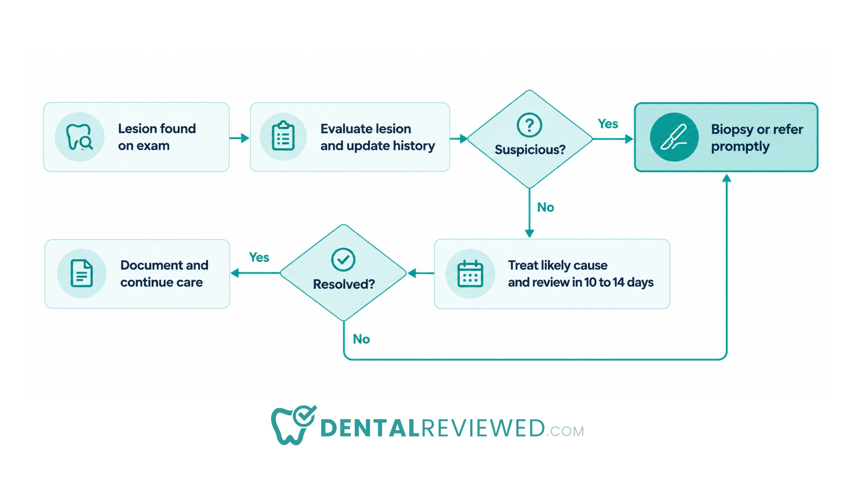
The pathway has one exit that skips biopsy – a lesion that heals once its cause is removed. Every other route ends at biopsy or referral.
What Does The ADA Say About Oral Cancer Screening Devices?
The guideline weighed four adjunct categories against one question. Should the test decide whether a patient needs biopsy or referral? The ADA announcement answers no for routine use, with one narrow cytology exception.
Method | Routine use to decide on biopsy? | Role under the 2026 guideline | Main limitation |
|---|---|---|---|
Clinical examination | Yes, for all adults | Foundation of early detection | Depends on consistent technique |
Biopsy with histopathology | When indicated | Definitive diagnosis | Invasive, may need referral |
Cytology | No | Option when biopsy is not possible, advisable, or indicated | A negative result does not rule out disease |
Vital staining | No | Not recommended for biopsy decisions or screening | Evidence does not support routine use |
Light-based adjuncts | No | Not recommended for biopsy decisions or screening | Evidence does not support routine use |
Salivary adjuncts | No | Research settings only | Not ready for clinical decisions |
The recommendations apply to two groups. One is adults with clinically evident lesions. The other is asymptomatic adults with no visible lesion. Neither group should receive adjunct testing as routine screening.
Cytology
Cytology collects loose cells from a lesion surface, usually with a brush. A laboratory then examines the cells for atypia.
The guideline does not recommend routine cytology to decide on biopsy or referral. It allows cytology in limited cases when biopsy is not possible, advisable, or indicated. Examples include a medically fragile patient or one who declines a scalpel procedure. A brush sample reaches surface cells only. Dysplasia deeper in the epithelium can be missed. A negative result therefore never closes the case.
Vital Staining
Vital stains such as toluidine blue bind to tissue with high nucleic acid content. Suspicious areas take up the dye and appear darker.
The guideline recommends against vital staining to decide on biopsy or referral. It also advises against staining to screen adults without visible abnormalities. Inflamed and ulcerated tissue can retain dye as well. A stained traumatic ulcer can look alarming and still be benign.
Light-Based Adjuncts
Light-based devices use autofluorescence or tissue reflectance to show changes in the mucosa. Healthy tissue fluoresces in a typical pattern, and some lesions lose that pattern.
The guideline generally recommends against routine use of these devices for biopsy decisions or screening. Loss of fluorescence occurs in inflammation and pigmented tissue too, so the signal lacks specificity. A device can also miss a lesion a careful palpation would catch.
Salivary Adjuncts
Salivary tests look for molecular biomarkers linked to oral cancer in a saliva sample. The appeal is a noninvasive test that needs no visible lesion.
The guideline recommends salivary adjuncts only in research settings. Validation data do not yet support their use in everyday clinical decisions. A practice offering these tests outside a study would be acting beyond the current ADA position.
Do Oral Cancer Screening Devices Still Have A Place?
The guideline narrows the role of adjuncts. Adjuncts keep a limited role. Reading the wording closely helps practices that already own a device.
The recommendations target one decision: whether a patient needs biopsy or referral. On that point, current evidence does not support routine adjunct use. Several recommendations are worded as "generally" against routine use. That phrasing signals limits in the evidence, and a living guideline can change once stronger studies appear.
How Can A Practice Use A Device It Already Owns?
A device can still add to a visit, provided it never overrides the exam. The safest rule is simple: no adjunct result can cancel an indicated biopsy or referral.
With a light-based device, a positive finding is a prompt to look again. A negative finding changes nothing. Clinical photographs of the same site, taken at each visit, offer a more useful record for the 10- to 14-day review.
What Should A Practice Ask Before Buying One?
New dental diagnostic tools reach the market every year, including AI tools that analyze lesion photographs. The guideline gives buyers a clear test for each one. The questions below follow from it:
Does the vendor claim the device decides on biopsy or referral?
Do published studies report results in a general dental population?
How often does the device flag inflamed or traumatic lesions?
Will a negative result tempt staff to skip a review visit?
A device that fails these questions adds cost and false reassurance. The living format means answers may change. Check the guideline page before any purchase.
What Should General Dentists Change In Practice?
Finding a lesion is only the first step. The guideline's real demand is a system that carries every lesion to a diagnosis.
Most delays happen between visits. A patient leaves with a vague plan to "keep an eye on it." The next look then comes at a six-month recall. The steps below close that path:
Use one exam sequence for every adult, at every visit type
Record lesion site, size in millimeters, color, texture, and duration
Photograph the lesion with a scale for side-by-side review
Note tobacco, alcohol, and other risk factors in the social history
Book the 10 to 14 day review before the patient leaves
Flag open lesions on a tracking list checked each week
Keep named referral contacts for oral surgery and oral medicine
Confirm receipt of every pathology report and file it
Each charted lesion should also appear on the dental treatment plan as an open diagnostic item. That keeps it visible to every provider who opens the record.
How Should Practices Handle the 10- to 14-Day Follow-Up?
The review visit is a diagnostic appointment. It has a different purpose from a routine recall.
Dr. Shelley Olson chairs the ADA Council on Dental Benefit Programs. She describes this visit as a medically necessary continuation of diagnosis. She advises coding it by the services performed and documented, regardless of prior exam dates. A refresher on dental codes helps the front desk file it correctly. The ADA also calls on payers to cover these follow-up visits, biopsies, and pathology services.
At the review, the clinician compares the lesion against the baseline record and photographs. A healed lesion closes the item. An unchanged, larger, or undiagnosed lesion moves straight to biopsy or referral that day.
How Should Dentists Talk To Patients About A Lesion?
Patients who understand the reason for a review return for it. Clear language works better than reassurance.
Explain that most mouth lesions are harmless and that the review confirms healing. State the date of the next visit and what will happen if the lesion remains. Avoid the word "cancer" until there is a reason to use it. Avoid promising that a lesion is benign. Tips on patient communication apply directly to these conversations.
Bottom Line
The 2026 ADA oral cancer guidelines point dentists toward a reliable clinical process. A thorough exam, a firm review date, and a timely biopsy or referral carry early detection. Adjuncts can sit beside that process in narrow roles. None of them can cancel a biopsy the clinical picture calls for. The practical test for any practice is simple. Does every suspicious lesion reach a definitive diagnosis on time?
Frequently Asked Questions
What are the new ADA oral cancer screening guidelines?
The 2026 living guideline calls for a full extraoral and intraoral exam on every adult. Suspicious lesions need prompt biopsy or referral. Lesions unresolved after 10 to 14 days need the same. The guideline advises against routine cytology, vital staining, light-based, and salivary adjuncts for biopsy decisions.
How often should dentists perform oral cancer screening?
The ADA recommends a comprehensive exam for all adults at new-patient, recall, and emergency visits. Each exam should include updated medical, social, and dental histories.
What does an oral cancer examination include?
It includes a visual and tactile check of the face, lips, neck, and lymph nodes. Inside the mouth, it covers the mucosa, gingiva, all tongue surfaces, floor of the mouth, palate, and oropharynx.
When should a dentist biopsy an oral lesion?
A dentist should biopsy or refer any suspicious lesion promptly. The same applies to a lesion that persists 10 to 14 days after evaluation without a definitive diagnosis.