Dental Diagnostic Tools: Technology Transforming Oral Care
Modern dentistry has witnessed a remarkable transformation in how professionals detect, diagnose, and treat oral health conditions. Advanced diagnostic technology enables more...
Written by Marcus Hale
Read time: 11 min read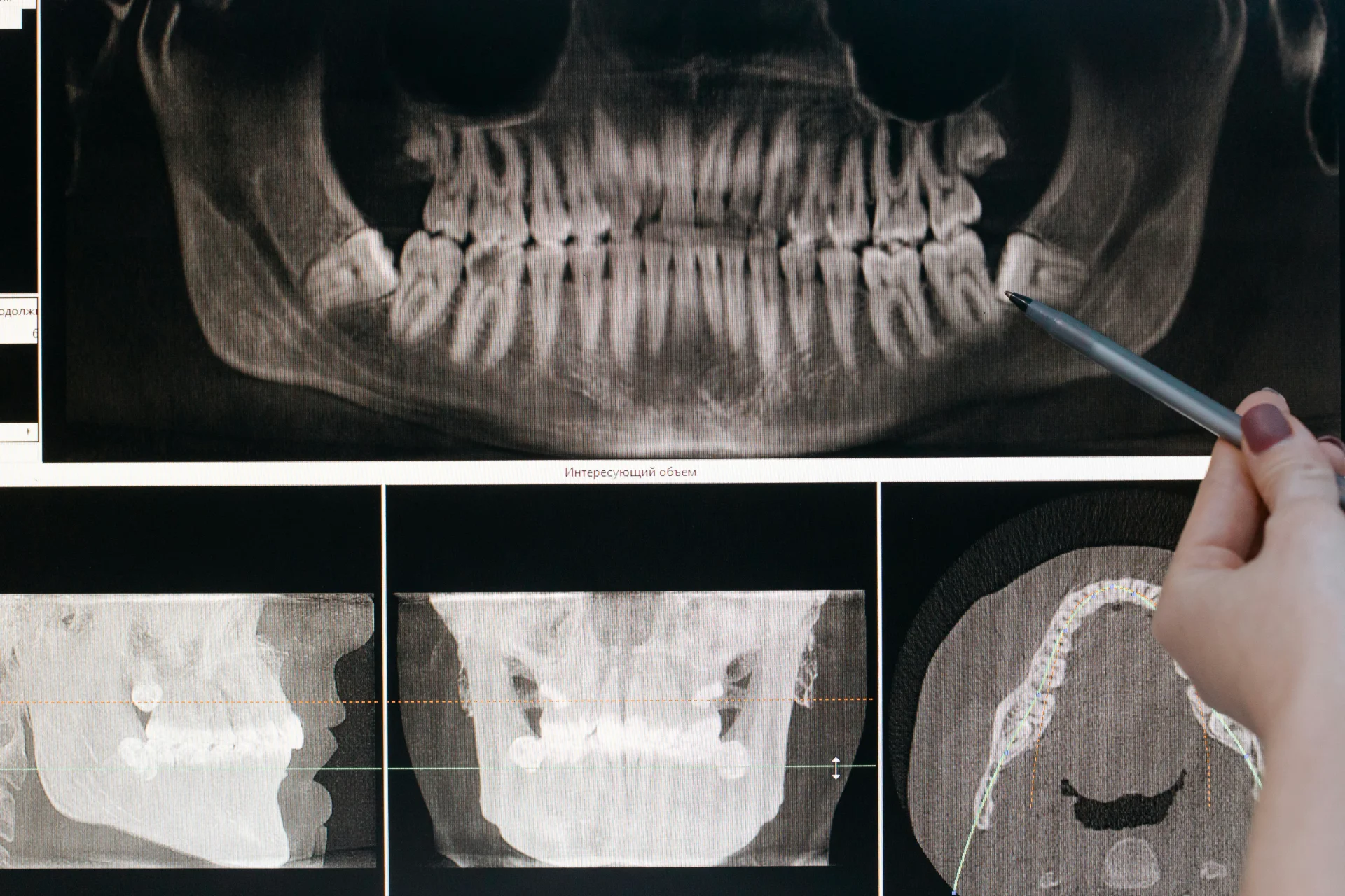
Modern dentistry has witnessed a remarkable transformation in how professionals detect, diagnose, and treat oral health conditions. Advanced diagnostic technology enables more precise diagnoses, personalized treatments, and enhanced patient experiences. The evolution from basic examination instruments to sophisticated digital systems has revolutionized patient care, making dental diagnostic tools more accurate, efficient, and comfortable than ever before.
Today's dental practices blend traditional examination methods with cutting-edge technology to provide comprehensive patient assessments. From handheld mirrors that have served dentists for over a century to artificial intelligence systems that can detect cavities invisible to the human eye, the range of available diagnostic equipment continues to expand. These innovations enable practitioners to identify oral health issues at their earliest stages, when treatment options are less invasive and more successful.
The integration of digital imaging, laser technology, and computer-aided systems has fundamentally changed how dental professionals approach diagnosis. Patients benefit from reduced radiation exposure, shorter appointment times, and clearer explanations of their oral health status through visual demonstrations. Meanwhile, dental practitioners gain access to more detailed information, improved accuracy, and better documentation capabilities that enhance treatment planning and outcomes.
This comprehensive guide explores the full spectrum of dental diagnostic tools available to modern practices. We will examine traditional instruments that remain essential to dental examinations, advanced imaging technologies that reveal hidden pathology, specialized equipment for detecting specific conditions, and emerging innovations that promise to further transform oral healthcare. Whether you are a dental professional evaluating equipment options, a student learning about diagnostic methods, or a patient curious about the technology used during your appointments, this guide provides the detailed information you need.
Essential Traditional Dental Diagnostic Tools
Despite remarkable technological advances, fundamental diagnostic instruments continue to play a crucial role in dental examinations. These time-tested tools provide tactile feedback and visual information that remain valuable alongside digital innovations. Traditional examination tools offer immediate, cost-effective diagnostic capabilities that require minimal maintenance and no specialized training beyond standard dental education.
Dental Mirrors and Explorers
The dental mouth mirror represents one of the most versatile instruments in any practitioner's toolkit. This simple device allows dentists to visualize areas of the oral cavity that would otherwise remain hidden from direct view, including the lingual surfaces of teeth, the posterior regions of the mouth, and the soft tissue structures. Modern dental mirrors come in various sizes and angles, with front-surface mirrors providing clearer, distortion-free images compared to traditional back-surface versions. Some contemporary designs incorporate LED lighting to illuminate examination areas more effectively.
Dental explorers, particularly the shepherd's hook or sickle-shaped varieties, enable practitioners to detect surface irregularities, assess restoration margins, and identify calculus deposits. While laser cavity detection systems have reduced reliance on explorers for diagnosing carious lesions, these instruments remain valuable for examining restoration quality and detecting structural abnormalities. The tactile sensation provided through careful exploration gives practitioners information about tooth structure integrity that visual inspection alone cannot reveal.
Contemporary dental practice recognizes that aggressive probing with explorers can potentially damage demineralized enamel in its earliest stages. Therefore, many practitioners now use these instruments with lighter pressure, focusing on areas where visual examination or other diagnostic methods suggest potential problems. This refined approach balances the benefits of tactile examination with the goal of preserving tooth structure whenever possible.
Periodontal Probes
Periodontal probes serve as the primary tool for assessing gum health and diagnosing periodontal disease. These calibrated instruments measure pocket depth, the space between the gum tissue and the tooth surface, which indicates the level of attachment loss associated with periodontal conditions. Standard probes feature millimeter markings that allow practitioners to record measurements at multiple points around each tooth, creating a comprehensive map of periodontal health.
According to the Centers for Disease Control and Prevention, periodontal disease affects a significant portion of the adult population. Regular probing during dental examinations enables early detection of gum disease when it remains most treatable. Different probe designs serve specific purposes, with Williams probes offering clear millimeter markings at 1, 2, 3, 5, 7, 8, 9, and 10 millimeters, while WHO probes include a colored band between 3.5 and 5.5 millimeters for quick screening.
Proper probing technique requires careful insertion of the instrument parallel to the tooth surface, gentle advancement until resistance indicates the base of the pocket, and systematic recording of measurements around the circumference of each tooth. The process provides essential baseline data for monitoring periodontal health over time and evaluating the effectiveness of treatment interventions. While electronic probes now offer automated measurement capabilities, traditional manual probes remain widely used due to their reliability, affordability, and the tactile feedback they provide.
Articulating Paper and Bite Registration Tools
Articulating paper helps dental professionals assess occlusion, the way upper and lower teeth contact during biting and chewing movements. This thin, color-coated paper transfers ink marks to tooth surfaces when patients bite down, revealing high spots, premature contacts, and occlusal patterns. The ability to visualize these contact points guides adjustments to restorations, orthodontic appliances, and natural tooth surfaces to achieve optimal occlusion.
Modern articulating papers come in various thicknesses and colors, allowing practitioners to differentiate between static contacts and dynamic movements. Red and blue papers used sequentially can distinguish between different phases of occlusion, while paper thickness affects the sensitivity of the marking process. Digital alternatives now exist, but paper remains popular due to its immediate results, low cost, and universal availability.
Bite registration materials complement articulating paper analysis, providing three-dimensional records of how teeth fit together. These materials, ranging from rigid waxes to flexible silicones, capture the spatial relationship between dental arches. The resulting registrations guide laboratory technicians in fabricating restorations, aid in diagnosing temporomandibular joint disorders, and document occlusal relationships for treatment planning purposes.
Advanced Dental Imaging Systems for Accurate Diagnosis
Digital imaging technology has revolutionized diagnostic capabilities in dentistry, providing clearer visualization of dental structures, reduced radiation exposure, and enhanced ability to detect problems at their earliest stages. These systems generate detailed images that can be immediately viewed, manipulated, and shared, transforming how dental professionals diagnose conditions and communicate with patients.
Digital Radiography (X-rays)
Digital dental X-rays have largely replaced traditional film-based radiography in modern practices, offering numerous advantages that enhance both diagnostic quality and patient safety. Digital imaging provides an opportunity to further reduce the radiation dose by 40 to 60 percent compared to conventional film methods. This significant reduction addresses long-standing concerns about cumulative radiation effects while maintaining diagnostic image quality.
Intraoral digital sensors capture X-ray images instantly, displaying them on computer screens within seconds of exposure. This immediate feedback allows practitioners to verify image quality during the appointment, eliminating the need for retakes due to positioning errors or processing problems common with film. The sensors connect directly to practice management software, automatically attaching images to patient records and facilitating comparisons with previous radiographs to track changes over time.
Panoramic digital X-rays provide a comprehensive view of all teeth, jaws, sinuses, and surrounding structures in a single image. These extraoral radiographs serve multiple purposes, including screening for impacted teeth, evaluating jaw development, detecting cysts or tumors, and planning complex treatments such as implant placement or orthodontics. Modern panoramic units often include additional imaging modes such as cephalometric views for orthodontic analysis and TMJ-specific projections.
Image enhancement tools available with digital radiography extend diagnostic capabilities beyond what film could offer. Practitioners can adjust contrast and brightness to better visualize specific structures, magnify areas of interest for detailed examination, apply false color to highlight density variations, and measure distances or angles directly on screen. These digital manipulation capabilities improve accuracy in detecting early decay, assessing bone levels, and planning precise treatments.
Integration with practice management software streamlines workflow and enhances patient communication. Digital images can be displayed chairside on large monitors, allowing patients to see what the dentist sees and better understand their oral health status. The ability to email images to specialists, insurance companies, or other healthcare providers facilitates collaborative care and expedites authorization processes. Cloud storage and automatic backup systems protect valuable diagnostic records while enabling access from multiple locations.
Cone Beam Computed Tomography (CBCT)
Cone beam computed tomography represents a major advancement in three-dimensional dental imaging, providing detailed volumetric data that two-dimensional radiographs cannot capture. CBCT scanners rotate around the patient's head, capturing hundreds of images from different angles that computer software assembles into a 3D model of dental and facial structures. This technology has become increasingly important for complex diagnostic scenarios and treatment planning.
Implant dentistry has particularly benefited from CBCT imaging capabilities. The technology allows precise measurement of available bone height and width, identification of vital structures such as nerves and sinuses, assessment of bone density, and virtual placement of implants before surgery begins. This comprehensive planning reduces surgical complications, improves implant positioning for optimal function and aesthetics, and enables the fabrication of surgical guides that translate virtual plans to actual procedures with remarkable accuracy.
Endodontic applications of CBCT include detecting accessory canals that might harbor infection, identifying root fractures that can be difficult to see on conventional radiographs, evaluating periapical pathology in three dimensions, and assessing complex root anatomy before treatment. Research demonstrates that CBCT imaging can reveal details missed on two-dimensional films, particularly in cases involving extra canals or unusual root configurations.
Orthodontic treatment planning utilizes CBCT data to analyze airway dimensions, assess impacted teeth positions, evaluate jaw relationships in all three planes of space, and plan skeletal anchorage device placement. The ability to visualize the complete craniofacial complex helps orthodontists make more informed decisions about treatment approaches and predict outcomes more accurately. Some practices now use CBCT scans as part of comprehensive orthodontic records for complex cases.
While CBCT provides exceptional diagnostic information, appropriate use guidelines recommend limiting scans to situations where the three-dimensional data will meaningfully influence treatment decisions. The radiation dose from CBCT varies depending on the field of view and resolution settings, but typically exceeds that of conventional dental radiographs. Evidence-based selection criteria ensure that the benefits of CBCT imaging justify the radiation exposure for each individual patient.
Intraoral Scanners
Intraoral scanners have transformed the process of capturing dental impressions, replacing uncomfortable impression materials with digital scanning technology that creates precise three-dimensional models of dental arches. These handheld devices use optical imaging to capture thousands of data points per second, assembling them into detailed digital replicas of teeth and soft tissues. The technology has improved patient comfort, increased accuracy, and streamlined workflows between dental offices and laboratories.
Modern intraoral scanners employ various imaging technologies, including confocal microscopy, structured light projection, and active triangulation, each with specific advantages for different clinical situations. The scanning process involves slowly moving the scanner tip over tooth surfaces while the device captures overlapping images that the software stitches into a complete model. Most systems provide real-time visualization, allowing practitioners to identify areas needing additional coverage and ensuring scan completeness before dismissing the patient.
CAD/CAM integration represents a major advantage of digital impression technology. Scans can be immediately transmitted to milling machines for same-day restorations, sent digitally to laboratories, eliminating shipping delays, archived for future reference or comparison, and used with virtual design software for treatment planning. This digital workflow reduces turnaround time for restorations, minimizes opportunities for dimensional distortion, and creates a permanent record that traditional impressions cannot provide.
Orthodontic applications of intraoral scanning include treatment planning with clear aligner systems, fabricating retainers and other appliances, monitoring tooth movement during treatment, and maintaining digital records that occupy no physical storage space. The ability to capture multiple scans over time allows precise measurement of tooth movement and comparison of treatment progress against predicted outcomes. Some orthodontic practices have completely eliminated traditional impression materials, relying exclusively on digital scanning for all appliance fabrication.
Patient acceptance of intraoral scanning significantly exceeds that of conventional impression taking. The elimination of impression trays and materials reduces gagging, allows normal breathing during the procedure, provides a more comfortable experience overall, and gives patients the ability to see their scan results immediately. These factors contribute to improved patient satisfaction and can enhance case acceptance for recommended treatments.
Laser and Light-Based Dental Diagnostic Equipment
Optical diagnostic technologies harness the properties of light and lasers to detect oral health problems that might escape detection through visual examination or traditional radiography. These non-invasive tools provide objective measurements and real-time visualization that enhance diagnostic accuracy while increasing patient comfort and acceptance.
Laser Cavity Detection Devices
Laser fluorescence cavity detection systems, such as DIAGNOdent and similar devices, use specific wavelengths of light to identify changes in tooth structure associated with early decay. When laser light illuminates a tooth, healthy enamel exhibits minimal fluorescence, while demineralized areas and bacterial byproducts produce measurable fluorescence that the device quantifies with numeric readings. Higher numbers indicate greater levels of demineralization or carious activity, helping practitioners identify problem areas that appear clinically sound or show only subtle visual changes.
The technology excels at detecting occlusal decay in the deep pits and fissures of posterior teeth, where traditional explorers and radiographs often fail to reveal early lesions. Laser fluorescence can identify demineralization before cavitation occurs, presenting opportunities for remineralization treatments rather than restorative intervention. This early detection capability aligns with minimally invasive dentistry principles that prioritize tooth preservation.
Smooth surface decay detection represents another important application of laser fluorescence technology. The devices can assess interproximal surfaces when used with specialized tips designed to access contact areas between teeth. While bitewing radiographs remain the standard for detecting interproximal decay, laser fluorescence offers a radiation-free complement that can be used more frequently for monitoring suspicious areas or tracking remineralization efforts.
Clinical protocols for laser cavity detection typically involve establishing baseline readings for all teeth and using these measurements to identify surfaces requiring closer evaluation. Readings above established thresholds indicate increased risk of carious lesions, prompting additional diagnostic steps such as radiographic examination or ongoing monitoring. The objective nature of the numeric readings reduces subjectivity in decay detection compared to purely visual and tactile methods.
Validation studies examining the accuracy of laser fluorescence systems show sensitivity and specificity values that vary depending on the clinical situation, tooth surface, and disease stage. The technology performs best when used as an adjunct to comprehensive clinical examination rather than as a standalone diagnostic method. Integration with visual inspection, radiography, and patient risk assessment provides the most complete picture of carious disease activity.
Intraoral Cameras
Intraoral cameras bring unprecedented visual documentation and patient education capabilities to dental practices. These compact, pen-sized or wand-shaped devices capture high-resolution images and video of oral structures, displaying them immediately on computer monitors or chairside screens. The magnification and illumination provided reveal details that patients could never see in a mirror, transforming abstract diagnoses into concrete visual evidence that enhances understanding and treatment acceptance.
The cameras typically feature built-in LED lighting, autofocus capabilities, and interfaces that integrate with practice management software for automatic image capture and storage. Wireless models eliminate cable management issues and allow more flexible positioning during examination. Some advanced systems include features such as fluorescence modes that highlight plaque or carious lesions, polarization filters that reduce glare from wet surfaces, and measurement tools that enable dimensional analysis directly from captured images.
Patient education represents one of the most valuable applications of intraoral cameras. When patients can see a cracked tooth, recurrent decay around a filling, or inflammation in their gum tissue, they develop a better understanding of their oral health status and the rationale for recommended treatments. This visual confirmation addresses skepticism about diagnoses, reduces the "just trust me" dynamic that can create resistance, and empowers patients to participate actively in treatment decisions.
Documentation capabilities of intraoral cameras extend beyond patient records to include insurance claim support, legal protection, and collaborative care communication. Before-and-after images demonstrate treatment outcomes, time-sequenced photos track the progression of conditions, and annotated images can be sent to specialists along with referrals. Many insurance companies now request photographic documentation for certain procedures, making intraoral cameras increasingly essential practice equipment.
Integration with digital systems allows intraoral camera images to be stored alongside radiographs, treatment notes, and other patient data in unified electronic health records. This consolidation simplifies record management, enables quick retrieval during appointments, and facilitates comprehensive documentation that meets regulatory requirements. Cloud-based storage systems ensure images remain accessible even if local hardware fails while enabling multi-location practices to share patient information seamlessly.
Caries Detection Lights and Transillumination
Transillumination technology uses fiber-optic light to illuminate teeth from various angles, revealing cracks, decay, and structural defects that appear as dark shadows against the otherwise translucent tooth structure. Systems like CariVu and others in this category offer radiation-free cavity detection that can be performed as frequently as needed without cumulative exposure concerns. The technology proves particularly effective for detecting interproximal caries without the need for bitewing radiographs.
The physics of transillumination relies on the light scattering properties of tooth structure. Healthy enamel and dentin transmit light relatively uniformly, creating a glowing appearance when illuminated. Demineralized areas, carious lesions, and cracks scatter light differently, appearing as dark regions in the otherwise bright tooth. This contrast makes pathology readily apparent, even when positioned at locations difficult to visualize through traditional methods.
Crack detection capabilities distinguish transillumination from most other diagnostic technologies. Craze lines in enamel, fractured cusps, and split teeth create distinct shadow patterns when transilluminated that might be invisible during visual examination. Early identification of cracks allows protective treatments such as crowns before the tooth fractures completely, potentially saving teeth that would otherwise require extraction.
Clinical protocols often incorporate transillumination as part of comprehensive examinations, particularly for patients at high risk for caries or with challenging anatomical features that complicate traditional radiography. The immediate visual feedback allows practitioners to show patients areas of concern during the same appointment when they are detected, facilitating timely treatment discussions. The ability to capture and store transillumination images creates documentation that can be referenced during future appointments to track whether suspicious areas have progressed.
Limitations of transillumination include reduced effectiveness in teeth with extensive restorations, difficulty assessing occlusal surfaces where the complex anatomy creates shadows unrelated to pathology, and potential for false positives in areas where anatomical variations create light scattering patterns. Understanding these constraints allows practitioners to use the technology appropriately as one component of a comprehensive diagnostic assessment rather than relying on it exclusively.
Specialized Tools for Gum and Soft Tissue Diagnosis
Periodontal and oral soft tissue conditions require specific diagnostic approaches that differ from those used for hard tissue assessment. Advanced technologies in this category enable early detection of gum disease, screening for oral cancer, and assessment of salivary factors that influence oral health.
Electronic Periodontal Probes
Electronic periodontal probes automate the measurement and recording of pocket depths, attachment levels, and other periodontal parameters. These computer-connected instruments use constant force mechanisms to standardize probing pressure, reducing the variability inherent in the manual probing technique. Measurements are instantly recorded in the patient's electronic chart, often accompanied by voice annotations that capture clinical observations during the examination.
The consistency provided through electronic probing enhances the reliability of periodontal assessments over time. Manual probing force can vary significantly between examiners and even between different measurements from the same examiner, potentially affecting the accuracy of readings used to monitor disease progression or treatment response. Electronic systems apply predetermined force levels, eliminating this variable and enabling more meaningful comparison of sequential examinations.
Efficiency gains from electronic probing include simultaneous measurement and documentation, reducing chair time compared to manual probing and separate charting. Voice-activated systems allow practitioners to narrate findings during examination, capturing detailed information about bleeding, suppuration, furcation involvement, and mobility without interrupting the examination flow. Integration with periodontal charting software generates comprehensive reports that can be printed for patient records or transmitted to specialists.
Patient communication benefits from electronic probing systems that can display graphic representations of periodontal health during the examination. Color-coded charts showing pocket depths, attachment loss, and bleeding sites provide visual evidence of periodontal disease that helps patients understand their condition and the importance of recommended treatments. Some systems track changes over time, creating visual progressions that demonstrate improvement or deterioration.
Oral Cancer Screening Devices
Oral cancer screening technologies supplement visual examination with adjunctive tools designed to identify abnormal tissue that might represent early malignant or premalignant changes. Devices such as VELscope use tissue fluorescence visualization, in which different wavelengths of light cause healthy tissue to fluoresce differently than abnormal tissue. Suspicious areas appear as dark patches against the bright green fluorescence of normal mucosa, prompting closer evaluation and possible biopsy.
The five-year survival rate for localized oral cancer is approximately 84%, declining to about 38% for cancers that have metastasized, according to data from the American Cancer Society. Regular screening, particularly for high-risk patients who use tobacco or consume alcohol heavily, represents an essential component of comprehensive dental care. Adjunctive screening devices enhance the ability to detect subtle changes that might be overlooked during visual examination alone.
Clinical protocols for adjunctive oral cancer screening typically involve initial visual examination under normal lighting, followed by assessment with the screening device. Areas that appear suspicious with either method warrant documentation, monitoring, or referral for biopsy, depending on clinical judgment and patient risk factors. The devices serve as adjuncts rather than replacements for thorough visual and palpation examination, which remain fundamental to oral cancer screening.
Integration into routine examinations has increased screening frequency and practitioner awareness of oral cancer risk. When screening devices are readily available and incorporated into standard examination protocols, dentists perform more comprehensive soft tissue assessments and discuss oral cancer risk factors with patients more regularly. This increased attention to soft tissue examination benefits patient outcomes through earlier detection and intervention.
Salivary Diagnostics
Salivary diagnostic testing represents an emerging field that analyzes saliva composition to assess oral disease risk, detect systemic conditions, and personalize preventive recommendations. Chairside tests can measure salivary flow rate, buffering capacity, and bacterial levels associated with caries risk. More advanced laboratory-based salivary testing can identify specific pathogens, inflammatory markers, and even genetic factors related to periodontal disease susceptibility.
Caries risk assessment through salivary analysis typically evaluates levels of Streptococcus mutans and Lactobacillus species, bacteria strongly associated with tooth decay. Patients with high bacterial counts receive intensified preventive recommendations, including more frequent professional cleanings, antimicrobial rinses, and dietary counseling. The objective data provided through testing helps justify aggressive preventive protocols and encourages patient compliance with recommendations.
Flow rate and buffering capacity testing identify patients with dry mouth conditions that increase caries risk dramatically. Reduced salivary flow can result from medications, medical treatments such as radiation therapy, or systemic conditions like Sjögren's syndrome. Identifying these patients allows implementation of targeted interventions, including prescription fluoride products, saliva substitutes, and modifications to treatment approaches that account for elevated risk.
Emerging applications of salivary diagnostics extend beyond oral health to include screening for systemic diseases, monitoring drug levels, and detecting biomarkers associated with various medical conditions. Research continues to explore saliva's potential as a diagnostic fluid that could rival blood testing for certain applications while offering a non-invasive, easily collected sample. As validation studies progress and test reliability improves, salivary diagnostics may become increasingly integrated into both dental and medical practice.
Artificial Intelligence in Dental Diagnostics
Artificial intelligence and machine learning technologies are beginning to transform dental diagnostics, offering computer-assisted detection systems that can identify pathology in radiographic images, predict treatment outcomes, and support clinical decision-making. AI can detect and quantify the radiographic extent of caries and has the capacity to autocorrect dental images, according to the American Dental Association.
AI-Powered Image Analysis
Computer vision algorithms applied to dental radiographs can identify carious lesions, periodontal bone loss, periapical pathology, and other conditions with accuracy that matches or exceeds human practitioners in some studies. The models exhibited a highest precision of 98% and 99.4% for tooth numbering and detection, respectively, thereby demonstrating the potential for AI models as a supplementary diagnostic tool in dental radiology. The AI systems analyze radiographic images, marking areas of concern and providing confidence scores for their findings.
Caries detection algorithms have shown particular promise in research studies, with some systems demonstrating sensitivity and specificity comparable to those of experienced dentists when analyzing bitewing radiographs. The AI can detect very early decay that appears as subtle density changes in enamel, potentially enabling intervention before traditional diagnostic thresholds are met. This enhanced detection capability aligns with preventive approaches that seek to arrest or reverse disease processes whenever possible.
Bone loss assessment through AI analysis of radiographs provides objective measurements of periodontal disease severity and progression. The algorithms can calculate bone levels around all teeth visible in an image, comparing measurements to age-appropriate norms and identifying areas of accelerated loss. Automated measurement eliminates the time-consuming manual process of marking bone levels and reduces inter-examiner variability that can affect assessment consistency.
The AI tool achieved a sensitivity of 91% and a specificity of 88% in identifying high-risk cases, according to research published in recent dental journals. Integration challenges include validating AI systems across diverse patient populations, addressing regulatory requirements for medical device software, and establishing workflows that incorporate computer-assisted detection efficiently. The technology works best when practitioners understand that AI serves as a diagnostic aid rather than a replacement for clinical judgment.
Digital Treatment Planning Software
Computer-aided treatment planning systems allow practitioners to visualize proposed treatments before beginning procedures, improving predictability and enabling patients to see expected outcomes. Smile design software can show how veneers, crowns, or orthodontic treatment will change a patient's appearance, facilitating informed consent and enhancing case acceptance. The ability to preview results addresses patient anxiety about major dental work and clarifies treatment objectives.
Orthodontic planning platforms analyze diagnostic records, including intraoral scans, photographs, and radiographs, to create virtual treatment simulations. The software can predict tooth movements, estimate treatment duration, and design clear aligner sequences or bracket positioning. Integration with manufacturing systems enables direct fabrication of customized appliances from the digital plan, streamlining the process from diagnosis through treatment delivery.
Implant planning software uses CBCT data to virtually place implants in optimal positions, considering available bone, adjacent structures, and prosthetic requirements. The software can design surgical guides that transfer the virtual plan to the surgical site with precision. This digital workflow reduces surgical time, improves accuracy, and enhances communication between the surgical and restorative teams when multiple practitioners are involved.
Practice Management Integration
Cloud-based diagnostic platforms enable seamless integration of diagnostic data from multiple sources into unified patient records accessible from any location. Radiographs, intraoral scans, photographs, and diagnostic test results can be viewed together during treatment planning, with the ability to compare current findings against historical data. This consolidation simplifies case management and reduces the risk of missing important diagnostic information scattered across different systems.
Interoperability standards such as DICOM for medical imaging allow diagnostic equipment from different manufacturers to communicate with practice management software and share data with specialists or referring practitioners. This connectivity eliminates manual data transfer processes, reduces opportunities for errors, and accelerates communication between healthcare providers. Patients benefit from more coordinated care when all treating practitioners can access relevant diagnostic information easily.
Remote consultation capabilities supported through cloud-based platforms enable specialists to review diagnostic images and provide guidance without requiring the patient to travel for an initial consultation. This teledentistry application proved particularly valuable during the pandemic and continues to offer convenience for patients and efficiency for practices. Secure messaging and annotation tools facilitate clear communication about diagnostic findings and treatment recommendations.
How to Choose The Right Diagnostic Equipment
Choosing appropriate diagnostic technology requires careful analysis of practice needs, patient demographics, clinical applications, and financial considerations. The abundance of available options can overwhelm practitioners, making a systematic evaluation process essential for making informed equipment decisions that enhance patient care while supporting practice success.
Practice size and patient volume directly influence equipment selection and return on investment calculations. High-volume practices may justify sophisticated diagnostic systems that would remain underutilized in smaller offices, while solo practitioners might prioritize versatile equipment that serves multiple diagnostic purposes. Understanding current patient flow and growth projections helps determine whether advanced technology will be used frequently enough to warrant the investment.
Budget considerations extend beyond initial purchase price to include installation costs, staff training expenses, ongoing maintenance fees, and supplies or disposables required for operation. Financing options such as equipment leasing or payment plans can make advanced technology accessible to practices with limited capital, though the total cost over time typically exceeds outright purchase prices. Comparing ownership costs across different systems and vendors ensures informed financial decisions.
Patient demographic analysis reveals whether specific diagnostic capabilities will significantly impact care delivery for your patient population. Practices serving many pediatric patients might prioritize radiation-free diagnostic tools, while those focused on implant or cosmetic dentistry require advanced imaging and planning software. Understanding the needs most common among your patients guides equipment selection toward tools that will be used regularly.
Training and learning curve factors affect how quickly new diagnostic equipment becomes productive in the practice. Systems with intuitive interfaces and comprehensive manufacturer training programs enable faster adoption and reduce frustration during the transition period. Evaluating the time required for staff to become proficient helps set realistic expectations and plan for temporary productivity decreases during implementation.
Technology upgrade versus replacement decisions should consider whether existing equipment remains functional and adequate for current needs or whether emerging capabilities justify replacement. Practices sometimes upgrade prematurely, replacing functional equipment with newer versions that provide marginal improvements. Conversely, continuing to use outdated technology when superior options exist can compromise diagnostic quality and practice competitiveness.
Vendor support and maintenance capabilities significantly impact long-term satisfaction with diagnostic equipment. Reliable technical support, prompt equipment service, and regular software updates ensure that systems remain functional and current throughout their useful lives. Evaluating vendor reputation, reading reviews from other practitioners, and understanding warranty terms helps avoid problematic relationships that can result in expensive downtime.
Integration with existing systems determines how seamlessly new diagnostic equipment will function within current practice workflows. Equipment that cannot communicate with practice management software creates inefficiencies and duplicate data entry that diminish the benefits of digital technology. Confirming compatibility before purchase prevents integration problems that can be difficult or expensive to resolve after installation.
Regulatory compliance requirements vary depending on equipment type and jurisdiction, with radiation-producing devices requiring specific safety protocols and regular inspections. Understanding these obligations and ensuring that your practice can meet them prevents regulatory violations that could result in fines or equipment restrictions. Some states mandate specific training or certification for operating certain types of diagnostic equipment.
How Modern Diagnostic Tools Improve Patient Care
The advancement of dental diagnostic technology has transformed patient care in ways that extend far beyond simply identifying oral health problems. These tools enhance nearly every aspect of the patient experience while enabling practitioners to deliver more precise, effective, and minimally invasive treatments.
Early detection represents perhaps the most significant benefit of modern diagnostic tools. Technologies such as laser fluorescence, transillumination, and high-resolution digital imaging reveal problems at stages when intervention remains straightforward and conservative. Catching decay before it requires extensive restoration, identifying periodontal disease before attachment loss becomes severe, and detecting oral cancer while it remains localized all dramatically improve treatment outcomes and long-term prognosis.
Accuracy improvements through objective measurements and enhanced visualization reduce diagnostic errors that can lead to unnecessary treatment or missed pathology. Multiple diagnostic technologies used together provide confirmation when findings align and prompt additional investigation when results conflict. This comprehensive approach minimizes both false positives that result in overtreatment and false negatives that allow disease to progress undetected.
Patient communication capabilities transform abstract diagnoses into concrete visual evidence that patients can understand and accept. Viewing their own radiographs, seeing magnified images of cracks or decay, and watching animations of planned treatments helps patients comprehend their oral health status and the rationale for recommended care. This improved understanding addresses skepticism, reduces anxiety about proposed treatments, and promotes informed decision-making.
Minimally invasive treatment opportunities arise when diagnostic tools detect problems early enough that conservative approaches remain viable. Remineralizing early carious lesions rather than placing fillings, using targeted antimicrobials for localized infections rather than systemic antibiotics, and monitoring suspicious lesions rather than performing unnecessary biopsies all preserve health while addressing problems effectively. These conservative approaches align with modern healthcare principles that prioritize preservation whenever possible.
Efficiency gains from digital diagnostic systems reduce appointment times, eliminate delays associated with film processing or impression shipping, and streamline workflows throughout the practice. Patients appreciate shorter appointments that accomplish more, while practices benefit from improved productivity that allows serving more patients or allocating more time to complex cases requiring extended attention.
Documentation quality achieved through digital systems creates comprehensive records that serve multiple purposes. Detailed diagnostic images support insurance claims, provide legal protection if treatment outcomes are questioned, facilitate communication with specialists and referring practitioners, and create baseline records against which future changes can be compared. The ease of duplicating and sharing digital records contrasts sharply with the limitations of physical films and paper documents.
Comfort improvements through less invasive diagnostic procedures enhance patient satisfaction and reduce anxiety about dental appointments. Replacing impression materials that can trigger gagging with digital scanning, using radiation-free diagnostic tools that can be applied repeatedly without concern, and employing painless screening technologies encourages patients to maintain regular examination schedules rather than avoiding dental care.
The Future of Dental Diagnostic Technology
Emerging innovations promise to further transform dental diagnostics over the coming years, with several technologies progressing from research laboratories toward clinical application. Understanding these developments helps practices prepare for the coming changes and recognize opportunities to adopt new capabilities as they become available.
Augmented reality applications could overlay diagnostic information directly onto the practitioner's view of the patient during examinations and procedures. Imagine wearing glasses that highlight areas of decay, display virtual guides for implant placement, or show the predicted results of treatment in real-time. This technology would merge digital treatment planning with direct visualization, potentially improving accuracy and efficiency simultaneously.
Molecular diagnostics may enable the detection of disease markers before any clinical signs become apparent. Salivary tests could identify genetic susceptibilities to periodontal disease, oral cancer risk factors, or metabolic conditions affecting oral health. This personalized risk assessment would allow truly preventive care targeted at each patient's specific vulnerabilities rather than applying standard recommendations to everyone.
Wearable oral health monitors represent an intriguing possibility for continuous tracking of oral conditions outside the dental office. Devices attached to teeth or incorporated into oral appliances could measure pH levels, detect bacterial activity, monitor bite forces, or track tooth movement during orthodontic treatment. Real-time data transmitted to practitioners would enable responsive care adjustments and early intervention when problems develop between appointments.
Teledentistry diagnostic tools continue evolving to support remote consultations and monitoring. Improved imaging devices designed for patient self-use, AI-powered analysis of patient-captured photos, and secure platforms for asynchronous consultation all expand access to dental care, particularly for underserved populations or patients with mobility limitations. The pandemic accelerated teledentistry adoption, and continued refinement will likely establish it as a standard care delivery mode.
Personalized medicine integration will increasingly connect oral health diagnostics with broader health monitoring and disease prevention. As understanding grows regarding links between oral health and systemic conditions, including cardiovascular disease, diabetes, and inflammatory disorders, diagnostic tools that assess these connections will become more prevalent. Dental practitioners may play expanding roles in overall health screening and disease risk assessment.
Blockchain technology could transform dental record management, creating secure, decentralized systems for storing and sharing diagnostic images and patient data. This approach would give patients greater control over their health information while ensuring that all treating practitioners can access necessary records. The technology could also support verification of credentials, authentication of treatment histories, and secure transmission of sensitive medical information.
Conclusion
Modern dental diagnostics have moved far beyond simple visual checks, becoming a cornerstone of precise, preventive, and patient-centered care. Traditional instruments combined with digital imaging, laser-based detection, AI-assisted analysis, and soft tissue screening technologies allow dental professionals to identify issues earlier, plan treatments more accurately, and communicate findings more clearly. The result is not just better diagnostics, but better outcomes – less invasive treatments, improved long-term oral health, and higher patient trust and satisfaction.
As technology continues to evolve, diagnostic tools will play an even greater role in shaping the future of oral healthcare. Practices that thoughtfully adopt and integrate these innovations are better positioned to deliver high-quality care, meet rising patient expectations, and stay competitive in an increasingly data-driven field. Ultimately, advanced diagnostics are not about replacing clinical expertise, but about enhancing it, giving dentists clearer insights, patients greater confidence, and oral healthcare a smarter, more preventive path forward.
Frequently Asked Questions
What are dental diagnostic tools?
Dental diagnostic tools are instruments and technologies used to examine the teeth, gums, and oral structures to detect disease, abnormalities, and risk factors. They range from traditional mirrors and probes to advanced digital imaging, lasers, and AI-powered software.
How do modern diagnostic tools improve dental care?
Modern tools improve accuracy, enable earlier detection, reduce radiation exposure, and support minimally invasive treatment. They also enhance patient understanding by providing clear visual evidence of oral health conditions.
Are advanced dental diagnostic technologies safe for patients?
Yes. Most modern diagnostic systems are designed to minimize risk, including low-radiation digital X-rays and non-invasive, light-based detection tools. When used appropriately, their benefits significantly outweigh potential risks.
Do all dental practices use the same diagnostic tools?
No. The tools used vary based on practice size, specialization, patient demographics, and budget. General practices may rely on digital X-rays and intraoral cameras, while implant, orthodontic, or specialist clinics often use CBCT, scanners, and AI-assisted planning tools.
Can dental problems really be detected before symptoms appear?
Yes. Technologies such as laser cavity detection, transillumination, digital imaging, and AI analysis can identify early-stage decay, gum disease, and structural changes before pain or visible damage occurs, allowing for more conservative treatment.