Dental Pulp Regeneration: New Study Finds A Potential Biological Switch
When deep decay reaches the pulp, the story often ends with a root canal and a tooth with no living tissue inside. Dental pulp regeneration tries to change that ending. A study in...
Written by Maren Solvik
Read time: 4 min read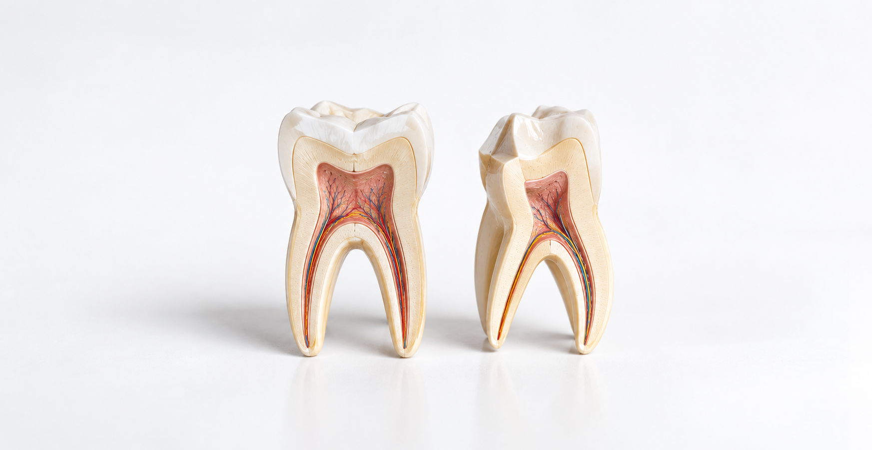
When deep decay reaches the pulp, the story often ends with a root canal and a tooth with no living tissue inside. Dental pulp regeneration tries to change that ending. A study in Experimental & Molecular Medicine, published October 6, adds an intriguing clue: a protein called fibronectin that seems to turn immune cells into allies of repair.
TL;DR
Researchers at Seoul National University traced how macrophages and pulp stromal cells communicate as decay deepens.
Fibronectin, a matrix protein, pushed macrophages toward a repair-friendly state that encourages new blood vessels.
In immature dog teeth, fibronectin thickened root dentin and raised apical closure from 22.7% to 59.1%.
The work is preclinical, so dentists cannot offer fibronectin pulp treatment to patients today.
Why Is Dental Pulp So Hard To Regenerate?
The pulp lives in an unusual place. It is soft, richly supplied tissue sealed inside a rigid shell of dentin, with one narrow doorway at the root tip for its blood vessels and nerves. Inflamed tissue elsewhere in the body can swell freely. Inflamed pulp has nowhere to go, so pressure builds against the walls and the blood supply struggles to keep up. That is a large part of why advanced pulpitis is so hard to reverse.
The same anatomy makes rebuilding pulp difficult. Getting new cells into a canal is only the first step. For that tissue to behave like real pulp, it needs:
Blood vessels to carry oxygen and nutrients
An organized extracellular matrix to hold everything in place
Odontoblasts lining the dentin wall, ready to make new dentin
Nerves and resident immune cells
A working connection with the surrounding dentin
Of these, the blood supply comes first. New tissue without vessels quickly starves, which is why angiogenesis, the growth of new blood vessels, sits at the heart of the new study.
What Did The New Study Set Out To Do?
Researchers at Seoul National University, led by Jin Man Kim, wanted to understand a conversation that happens inside every inflamed pulp. As decay advances, immune cells called macrophages pour into the tissue and press up against the pulp's own stromal cells. The team asked what those two cell types say to each other, and whether the exchange helps or hurts healing.
Their paper moves through several levels of evidence, and it helps to keep them apart:
Evidence level | What researchers did | What it shows |
|---|---|---|
Human tissue | Examined pulp from healthy teeth and teeth with moderate or severe caries | How macrophages change as decay advances |
Laboratory | Grew macrophages with dental pulp stromal cells and blocked key signals | How fibronectin drives the macrophage shift |
Single-cell data | Re-analyzed public human pulp datasets | Which cells signal to which as caries progresses |
Animal model | Treated immature dog teeth with fibronectin during revascularization | Whether the pathway improves regeneration in a living tooth |
Each layer answers a different question, and only the last one tests a treatment.
Can Immune Cells Help The Pulp Heal?
Macrophages have a reputation as the body's demolition crew. They swallow bacteria, clear dead cells, and sound the alarm that brings in more immune defenders. All of that is true, yet it leaves out half of their job. The same cells release growth factors, remodel damaged tissue, and guide the stem and stromal cells around them during healing.
What a macrophage does depends on the signals it receives. Researchers often sort these cells into two broad states: M1 cells that drive inflammation and M2 cells that support repair. Real macrophages sit along a spectrum, though the labels make it easier to see which way a population is leaning.
For endodontics, this reframes inflammation. The aim may be to steer the immune response at the right moment, so that cells already in the pulp start rebuilding it. A January 2026 laboratory study reached a related conclusion, finding that injured pulp fibroblasts help decide which state nearby macrophages adopt.
What Happened Inside Decayed Human Teeth?
The investigation started with real teeth. The team examined pulp from healthy teeth and from teeth with moderate or severe caries, staining it to locate macrophages and stromal cells.
In healthy pulp, macrophages were sparse and spread evenly through the tissue. As decay moved closer, they gathered beneath the infected dentin near the odontoblast layer. In severe caries, they flooded the pulp, packed most densely under the deepest part of the lesion. More telling was how often they were physically touching the pulp's stromal cells:
Caries stage | Macrophages in contact with stromal cells |
|---|---|
Healthy | 11.2% |
Moderate | 23.7% |
Severe | 48.7% |
Contact roughly doubled at each stage. To check whether the pattern held across the whole tissue, the researchers re-analyzed public single-cell datasets from human pulp. Immune cells rose from 1.5% of pulp cells in healthy teeth to 18.1% in severe caries, and endothelial cells, which line blood vessels, fell from 16.8% to 9.5%. The number of predicted conversations between cell types jumped from 278 to 2,470.
These observations describe what decay does to pulp, and they test no treatment. The single-cell samples were also small, with only one tooth each in the moderate and severe groups.
How Did Fibronectin Emerge As The Key Signal?
Fibronectin is one of the proteins that make up the extracellular matrix, the scaffolding between cells. During wound healing, it helps cells attach, move, and organize, and cells read it through surface receptors called integrins.
When the team grew macrophages alongside pulp stromal cells, they saw the stromal cells laying down fibronectin and the macrophages gripping it with an integrin called α5β1. They then tried to break that link in several ways:
An RGD peptide that blocks integrin binding reduced contact between the two cell types
Macrophages engineered to lack integrin α5 made fewer and shorter contacts
Growing macrophages on fibronectin raised pro-angiogenic chemokines in a dose-dependent way
Silencing fibronectin in the stromal cells lowered those same signals
Inside the macrophage, the message traveled through a transcription factor called NFκB. Inflammatory chemokines such as CCL2 and CCL3 dropped, and angiogenic CXC chemokines and VEGF-A climbed. More of the macrophages also carried CD206, a marker of the reparative M2 state.
The most interesting character in this story may be the macrophage itself. Inside inflamed pulp, it is usually treated as part of the damage. Given the right matrix signal, it appeared to switch sides and join the repair effort.
Why Do New Blood Vessels Matter So Much?
A living pulp is only as healthy as its blood supply. Vessels bring oxygen, nutrients, immune cells, and signaling molecules into the canal, and they carry metabolic waste back out. Regenerated tissue without that supply would struggle to survive for long.
To test whether the macrophage shift translated into vessel growth, the researchers bathed human endothelial cells in fluid collected from their co-cultures. Those cells formed more tube-like networks, the lab version of early blood vessels, than cells exposed to fluid from either cell type alone. They also died less often. Blocking integrins, the CXCR2 receptor, or VEGF receptors weakened both effects, which tied the vessel growth back to the fibronectin pathway.
What Happened When Researchers Added Fibronectin To Dog Teeth?
The final test moved from the dish to living teeth. Consider the kind of case regenerative endodontics was built for: a young permanent tooth whose pulp has died before the root finished growing. The root is short, its walls are thin, and the tip is still wide open. A conventional root canal can save that tooth, yet the root stops developing and stays prone to fracture.
The team recreated that situation in the immature premolars of three dogs, treating one side of the mouth and keeping the other as a control. They removed the pulp, disinfected each canal following American Association of Endodontists regenerative protocols, and then provoked bleeding into the canal to form a clot. Both sides received a collagen sponge and an MTA seal, and the fibronectin side also got a fibronectin rinse and a fibronectin-soaked sponge. Eight weeks later, they compared the results.
Outcome | Control roots (n = 22) | Fibronectin roots (n = 22) |
|---|---|---|
Mean root dentin area | 2.39 mm² | 3.40 mm² |
Roots with apical closure | 5 (22.7%) | 13 (59.1%) |
Canal tissue | Loose, amorphous fibrous tissue | Organized pulp–dentin-like tissue |
Blood vessels | Fewer | More, in apical and stromal regions |
Odontoblast-like layer | Not evident | Polarized cells positive for dentin sialoprotein |
Why Do Thicker Root Walls Matter?
Thin dentin walls are the weak point of an immature tooth with a dead pulp, and they leave those teeth prone to fracture. The fibronectin-treated roots ended up with about 40% more root dentin area, which points toward a sturdier root.
How Often Did The Root Tips Close?
Apical closure is a sign that the root kept developing. It happened in 13 of 22 fibronectin-treated roots compared with 5 of 22 controls. Some treated roots even formed the channel-like structures typical of a mature dog's root tip.
What Did The Tissue Inside The Canal Look Like?
Current regenerative procedures often fill the canal with loose fibrous tissue and scattered calcifications, and the control roots in this study looked much the same. The fibronectin-treated roots held something more organized: a layer of polarized odontoblast-like cells, dentinal tubule-like structures, and staining for dentin sialoprotein, a marker of dentin-forming cells. They also contained more reparative M2 macrophages, echoing what the team had seen in the dish.
What Can The Dog Results Not Tell Us?
These results come from three animals followed for eight weeks. The pulp in those teeth was healthy before it was removed under sterile conditions, so the canals never faced the bacteria and long-standing inflammation of a real decayed tooth. The study does not show that fibronectin can regenerate diseased pulp in people.
Is This The Same As Regrowing A Tooth?
No. Headlines often use "tooth regeneration" for several very different ideas, and it helps to separate them:
Concept | What it means | Status |
|---|---|---|
Enamel remineralization | Restoring mineral to early enamel lesions | Routine with fluoride and other agents |
Tertiary dentin | New dentin the pulp lays down after injury | Natural response |
Pulp regeneration | Restoring living tissue inside the tooth | Partly achieved in immature teeth |
Continued root development | Roots keep growing after treatment | Goal of regenerative endodontics |
Whole-tooth regeneration | Growing an entire new tooth | Experimental |
This study belongs to the pulp–dentin complex. Growing an entire new tooth is a separate field, explored in this look at whether teeth can regrow.
Can Dental Pulp Already Heal Itself?
Sometimes, and dentists rely on that ability every day. Healthy pulp responds to irritation by laying down tertiary dentin, a protective layer between itself and the threat. Whether an inflamed pulp can recover depends on:
How severe and long-lasting the inflammation is
How heavily bacteria have contaminated the pulp
How much healthy tissue remains
Whether the infection can be brought under control
The quality of the final seal
The blood supply and patient-specific factors
That natural capacity is what vital pulp therapy builds on. Indirect pulp treatment, direct pulp capping, partial pulpotomy, and full pulpotomy all aim to remove what is lost and protect what can still recover, often with calcium silicate materials such as Biodentine. Recent trials of partial pulpotomy in mature molars are covered in our pulpotomy vs. root canal analysis.
How Is Pulp Regeneration Different From A Root Canal?
A root canal removes diseased pulp, disinfects the canal system, and seals it with a filling material. It keeps the tooth in the mouth after the living tissue inside is gone, and it does that job very well. The trade-off is that the tooth can no longer form new dentin, and in a young tooth the root stops developing.
Regeneration aims for a different outcome, with living tissue back inside the canal. The main approaches line up like this:
Approach | Primary goal |
|---|---|
Root canal treatment | Keep the tooth after removing diseased pulp |
Vital pulp therapy | Preserve the remaining healthy pulp |
Regenerative endodontics | Encourage living tissue and continued root growth |
Experimental pulp regeneration | Recreate functional pulp–dentin tissue |
Root canal treatment remains a reliable choice when pulp cannot be saved, including when infection has spread and formed a tooth abscess.
Could This Help Patients Avoid Root Canals One Day?
Possibly, though not soon and not on the strength of this study alone. Vital pulp and regenerative approaches could gradually widen the range of teeth that keep living tissue. This paper adds one piece to the biological map those future therapies will need, and it does not establish fibronectin as a root canal alternative in humans.
The two lines of research answer different questions. Pulpotomy trials ask how far existing vital pulp therapy can go today. Studies like this one ask whether damaged pulp biology itself can be rebuilt.
Doesn't Regenerative Endodontics Already Exist?
It does, mainly for immature permanent teeth with necrotic pulp. Clinicians disinfect the canal, provoke bleeding to fill it with a blood clot, and seal it, hoping the root will keep growing and the tooth will heal. An expert consensus statement describes the current protocols.
Outcomes are less predictable than clinicians would like, as a 2024 network meta-analysis of these procedures shows. The authors of the new paper note that tissue growing inside treated canals is often fibrous connective tissue mixed with calcified deposits, and true odontoblasts forming tubular dentin are rarely seen. That shortfall is why researchers keep testing stem cells, scaffolds, growth factors and, now, immune signals.
Why Could Steering Macrophages Matter Beyond This Study?
For years, regeneration research has focused on three ingredients: the right cells, a scaffold to hold them, and signaling molecules to direct them. Injured tissue brings a fourth ingredient whether researchers plan for it or not, and that is an active immune environment.
This study suggests that the environment can be steered with something as ordinary as a matrix protein, which worked here by changing how resident immune cells behaved. The authors point out that fibronectin is a protein the body already makes, which may ease safety concerns compared with synthetic agents. If that holds up, future pulp therapies may be designed around immune signals as carefully as around scaffolds.
What Needs To Happen Before Patients Can Benefit?
The authors are candid that their dog study is a starting point. They call for work on delivery, dosing, long-term follow-up and, eventually, clinical trials. Several other hurdles follow from how the model was built, and from what makes a human tooth with deep decay so different from a freshly cleaned canal in a healthy young dog.
Is Fibronectin Safe Inside Human Pulp?
The authors argue that fibronectin should carry a low risk because the body already makes it. Blood plasma carries fibronectin, and pulp fibroblasts produce it constantly, as the team's own single-cell analysis showed. That is a reasonable starting assumption, though it falls short of safety data. A protein the body makes can still behave differently when applied at high concentration, in a new location, at a moment when the tissue is inflamed.
The mechanism adds a specific question. In the macrophages studied here, fibronectin worked through NFκB, a transcription factor better known for switching on inflammation. In this setting, the result was a pro-angiogenic, repair-friendly profile. Human studies would need to confirm that the same switch does not tip toward more inflammation in pulp that is already irritated, and that nearby tissues around the root tip respond well. Early safety work would also look for pain, swelling, unexpected calcification, and any immune reaction to the product itself.
How Would Dentists Deliver It?
In the dogs, fibronectin reached the canal in two ways. The final irrigation step soaked the canal in a 100 µg/ml fibronectin solution for 10 minutes, and a collagen sponge loaded with the same solution sat on top of the blood clot beneath the MTA seal. That is a practical research setup, and it shows how much of the effect depends on the carrier.
A clinical product would need to keep fibronectin in place and active for long enough to influence the cells moving into the canal. Researchers could tie it to an injectable hydrogel, build it into a scaffold, or blend it into a calcium silicate pulp-capping material for vital pulp therapy. Each option raises its own questions about release speed, shelf life, and how the material handles in a busy operatory. Decellularized pulp matrix hydrogels, which naturally contain fibronectin, are one related line of research already being tested in animals.
What Dose And Timing Work Best?
The study tested a single concentration delivered once, at the moment of revascularization. The laboratory work showed that macrophages raised their pro-angiogenic signals in a dose-dependent way as fibronectin levels rose, which suggests the dose matters. It does not tell us where the useful range ends or whether more fibronectin would keep helping.
Timing may matter just as much. Macrophages shift through different states as healing moves from early inflammation toward rebuilding. A signal that helps during the first days might be less useful, or even counterproductive, weeks later. Future studies will need to compare concentrations, contact times, and carrier materials, ideally while tracking how the macrophage population changes over time.
Will It Work In Infected Teeth?
This may be the biggest hurdle. The dog teeth started with healthy pulp, which was removed under sterile conditions before the canals were disinfected. Real patients arrive with something very different: bacteria and their toxins packed into dentinal tubules, a biofilm on the canal walls, and inflammation that may have simmered for months.
Regenerative procedures already struggle with this tension. Disinfection has to be strong enough to control infection, yet gentle enough to spare the stem cells from the root tip that the procedure relies on. Many current protocols favor lower-concentration sodium hypochlorite for that reason, while the dog study used a 5.25% solution in canals that held no infection. Any fibronectin-based approach will have to show that it still steers macrophages toward repair when bacterial signals are pushing them hard in the other direction.
Which Patients Would It Suit?
Pulp problems cover a wide range of clinical situations, and a treatment that helps one may not suit another. Reversible pulpitis, irreversible pulpitis, necrotic pulp in an immature tooth, and necrotic pulp in a fully formed tooth all start from different biology.
The dog model maps most closely onto immature permanent teeth with dead pulp, the group regenerative endodontics already treats. Those teeth have a wide-open root tip and a rich supply of stem cells in the tissue around it, which makes regrowth more realistic. Mature teeth have a narrow apical opening that limits how many cells and vessels can move in. A fibronectin-loaded capping material for vital pulp therapy would be a different product again, aimed at inflamed but living pulp. Each of these scenarios would need its own studies, with its own success criteria.
Does The New Tissue Work Like Real Pulp?
The fibronectin-treated roots looked impressive under the microscope, with odontoblast-like cells, tubule-like structures, and staining for dentin sialoprotein. Histology shows what tissue looks like at one moment, though, and pulp is judged by what it does over years. Future studies will need evidence on:
Blood flow, which clinicians can measure with tools such as laser Doppler flowmetry
Nerve supply and a reliable response to cold or electric pulp testing
Immune defense against later bacterial challenges
Odontoblast activity and continued dentin formation
Whether the canal stays open or gradually calcifies
How the tissue responds to a new injury or new decay
Current regenerative procedures show why these measures matter. A treated tooth can look healthy on a radiograph while the tissue inside behaves more like scar than pulp. Showing that fibronectin produces something closer to true pulp would require functional tests, long follow-up, and comparisons with untreated teeth.
Can It Be Made And Approved As A Dental Product?
The dogs received a research-grade fibronectin solution. A product for patients would need consistent, pharmaceutical-grade manufacturing, a stable formulation, and a clear regulatory pathway. Because it combines a biologic protein with a delivery material, it could face more scrutiny than a conventional dental cement, and cost will shape whether clinics can realistically use it.
How Will It Compare In Clinical Trials?
Eventually, fibronectin-enhanced procedures will need randomized trials against today's standard regenerative protocol, which relies on a blood clot alone. A useful trial would follow patients for several years and measure continued root development, tooth survival, healing at the root tip, and pulp sensibility. It would also need to report complications such as discoloration, canal calcification, and reinfection.
The dog study offers a useful template for what to measure, with root dentin area and apical closure as clear early signals. Human trials would need to show that those gains translate into teeth that last longer and function better, because that is the outcome patients and dentists care about.
What Does Today's Study Change?
Dentistry already knew that pulp cells can repair damage, that new blood vessels drive regeneration, and that macrophages flood inflamed pulp. What this study adds is a thread running through all three:
Caries injures the pulp
Pulp stromal cells lay down fibronectin
Macrophages grip it through integrin α5β1
Macrophages shift toward a pro-angiogenic state
New blood vessels form
Pulp–dentin regeneration improves in an animal model
The results remain preclinical, and fibronectin-based pulp regeneration is not an established human treatment. Dentists should not start using fibronectin because of this paper. The question it leaves open is whether steering this pathway can make pulp regeneration more predictable in people.
Bottom Line
Dental pulp regeneration took a step forward on October 6, 2026, when researchers showed how fibronectin can shift pulp macrophages toward a state that builds blood vessels. In immature dog teeth, the protein thickened root dentin and more than doubled the rate of apical closure. The findings are still preclinical, and vital pulp therapy and root canal treatment remain the evidence-based options for patients today.
Frequently Asked Questions
Can damaged dental pulp regenerate?
Pulp has some natural repair capacity, and regenerative endodontic procedures can encourage new tissue growth in selected immature teeth. Restoring pulp with its normal structure and function is still a research goal.
Can tooth pulp heal itself?
Sometimes. Mild inflammation can settle once the cause is removed, while a deep or long-standing infection can overwhelm the pulp's ability to recover.
Can an inflamed tooth nerve heal?
What people call the "nerve" is the pulp, which contains nerves, blood vessels, connective tissue, and cells. Reversible pulpitis can calm down once the cause is treated, and irreversible pulpitis usually needs a pulpotomy or root canal treatment.
What is dental pulp regeneration?
Dental pulp regeneration means restoring living, working tissue inside a tooth. Ideally, that tissue would include blood vessels, nerves, and odontoblasts able to form new dentin.
What is regenerative endodontics?
Regenerative endodontics covers procedures that encourage new tissue to grow inside the root canal. Dentists use them mainly for immature permanent teeth whose pulp has died.
Can dental pulp stem cells regrow a tooth?
No current clinical evidence shows that dental pulp stem cells can regrow an entire human tooth. Research focuses on regenerating tissue inside an existing tooth.
Can dentin regenerate?
The pulp–dentin complex can form tertiary dentin after injury, which adds a protective layer. That response cannot replace large amounts of lost tooth structure.