Enamel Hypoplasia: Causes, Symptoms, and Treatment Options
Enamel hypoplasia is a condition in which a tooth grows in with less enamel than it should have. It usually shows up as a small pit, a line across the tooth, or a tooth that seems...
Written by Rachel Thompson
Read time: 4 min read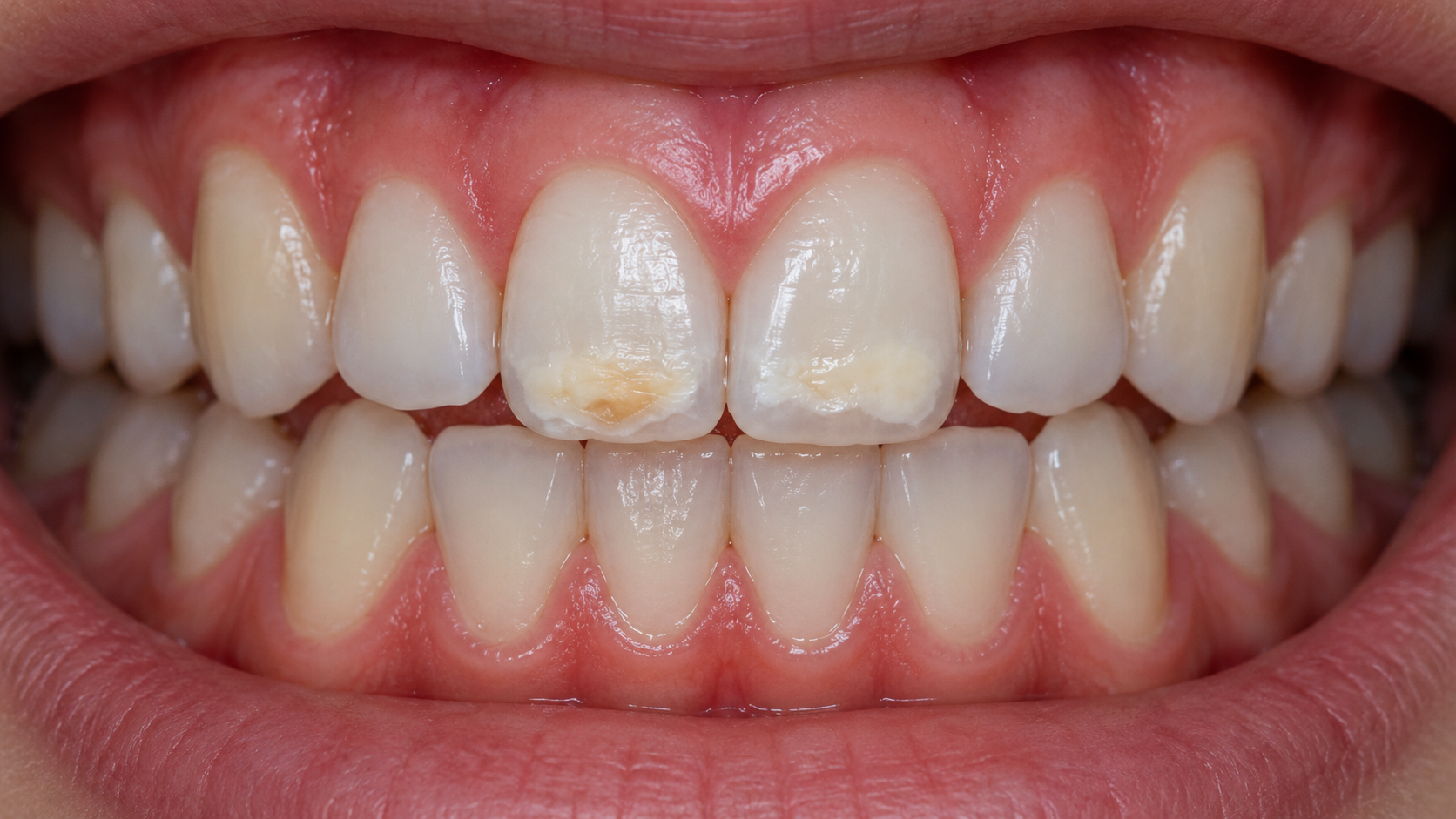
Enamel hypoplasia is a condition in which a tooth grows in with less enamel than it should have. It usually shows up as a small pit, a line across the tooth, or a tooth that seems to chip more easily than the rest. Each of those marks traces back to the time the tooth was still forming.
TL;DR
Enamel hypoplasia leaves teeth with thin, pitted, or missing enamel.
Dentists distinguish it from fluorosis, MIH, and early tooth decay.
Fluoride protects the remaining enamel but cannot replace what is missing.
Bonding, fillings, veneers, or crowns protect teeth with larger defects.
What Is Enamel Hypoplasia?
Picture a seven-year-old whose new front tooth has just come in. Mom notices a rough little dent near the edge and assumes it must have been chipped at school. At the dental visit, she learns the dent has been there all along. The tooth simply came in that way.
That is enamel hypoplasia in a nutshell. While a tooth is still tucked inside the jaw, specialized cells build its enamel layer by layer. If something interrupts those cells partway through the job, the enamel ends up thin in some spots or missing altogether. The tooth erupts with the defect already in place.
The catch is that those enamel-building cells disappear once the tooth comes through the gum. Bone can heal and skin can regrow, but enamel has no way to repair itself. A hypoplastic spot at age seven will look much the same at forty unless decay or wear changes it. The reassuring part is that the dentin and nerve underneath are usually perfectly healthy, and with sensible care these teeth can last a lifetime.
What Makes Hypoplasia Different From Stains?
The simplest way to put it is that a stain changes the color of a tooth, while hypoplasia changes its shape. That is why whitening can lift a coffee stain but leaves a hypoplastic pit looking exactly the same. When a dentist runs an instrument across the area, there is usually a small ledge where the surface drops away.
The two often show up together, which is where the confusion starts. A rough, pitted surface grabs pigment from coffee, tea, and red wine far more easily than smooth enamel does. Over the years, a hypoplastic spot slowly turns brown, and the patient sees a stain sitting on top of what is really a structural issue.
Enamel hypoplasia | Stain on normal enamel | |
|---|---|---|
Enamel thickness | Thin or missing in one area | Normal |
Surface texture | Rough, pitted, or grooved | Smooth |
When it appears | When the tooth erupts | Usually develops later |
After cleaning or whitening | Shape stays the same | Often lightens |
Where Does the Enamel Go?
With hypoplasia, the enamel never formed in the first place. The permanent front teeth and first molars build most of their enamel between birth and about age five, and that stretch of early childhood is when they are most vulnerable. A high fever, a serious infection, or a knock to the baby tooth above can make the enamel-building cells pause.
When growth picks back up, the tooth carries a permanent mark at the level that was forming at that moment. Depending on how long the pause lasted, it may look like a faint band, a few pits, or a bare patch.
This is a different process from acid erosion or tooth decay, both of which strip enamel away after a tooth is already in the mouth. Still, the two can meet in the same tooth. Pits and thin spots collect plaque, and with less enamel standing in the way, a cavity in a hypoplastic tooth can reach the softer dentin faster than usual.
What Does Enamel Hypoplasia Look Like?
Many people feel enamel hypoplasia before they ever see it. The tongue keeps finding a rough patch or a ridge that looks perfectly fine in the mirror. A closer look under good light usually reveals small pits, a line running across the tooth, or a flat, slightly yellow area where the surface seems to dip.
Just as telling is which teeth are affected and where the marks sit. A child with one rough front tooth and an otherwise perfect smile probably had something happen to that single tooth. A child with matching grooves at the same height on several teeth likely went through an illness while those teeth were forming together.
Pits, Grooves, and Missing Enamel
These three features are the most dependable signs of hypoplasia, since each one marks a spot where enamel stopped short of its full thickness. Pits tend to be small and rounded, sometimes alone and sometimes in clusters. Grooves run sideways across the tooth, roughly parallel to the gumline, like a faint line drawn with a ruler.
Where enamel is missing altogether, the surface looks flat or stepped, and the yellow dentin underneath starts to show through. When the whole enamel layer is thin, a tooth can look a little smaller than its neighbor or slightly glassy at the edges.
White, Yellow, or Brown Changes and Their Limits as Diagnostic Clues
Color changes often come along with hypoplasia, but on their own they are one of the weakest clues to go on. Yellow usually means dentin is peeking through thin enamel. Brown tends to build up over years as food and drink settle into a rough surface. White can sit right next to a defect or appear on a tooth with no structural problem at all.
The tricky part is that fluorosis, MIH, and early decay can all produce the very same shades. A smooth, chalky white patch is more likely to be weak enamel than missing enamel. A brown spot might be harmless staining inside a pit or the beginning of a cavity, and in a photo the two can look almost identical. That is why white spots and discoloration always deserve an exam before anyone settles on a cause.
Single-Tooth vs. Multiple-Tooth Patterns
The number and position of affected teeth give dentists a rough timeline to work with. Teeth that develop during the same months share the same risks, so their defects tend to show up together.
A single affected adult tooth usually points back to an injury or infection in the baby tooth that sat right above it. Defects lined up across several teeth suggest something that affected the whole body in early childhood, such as a long illness. When nearly every tooth is involved, in both the baby and adult sets, an inherited enamel disorder moves to the top of the list, and family history becomes one of the most useful questions in the room.
What Causes Enamel Hypoplasia?
Enamel hypoplasia develops when something interrupts enamel growth while a tooth is forming, and the timing decides which teeth end up marked. Baby teeth form mostly during pregnancy, so the mother's health and premature birth tend to show up on the primary teeth. The permanent front teeth and first molars form from birth to around age five, so illnesses and injuries in those early years leave their mark on the adult smile that appears much later.
In real life, the cause is often hard to pin down. Many children have more than one possible risk factor, and some have none that anyone can identify. Most of the links described below come from studies that show an association, which is not quite the same as proof.
Trauma or Infection Affecting a Baby Tooth
When only one adult tooth is affected, the story very often starts with the baby tooth that came before it. Dentists call this a Turner's tooth. The root of each baby tooth sits right on top of the adult tooth developing beneath it, separated by only a thin layer of bone.
Two scenarios account for most cases. In the first, a toddler takes a tumble, and a baby front tooth gets pushed up into the gum, bruising the adult tooth forming above it. In the second, a baby molar develops a deep cavity that turns into an abscess, and the infection spreads to the premolar growing underneath.
Either way, the consequence does not show up until years later, when the adult tooth erupts with a pit, a yellow-brown patch, or an unusual shape. This delayed effect is one of the less obvious reasons dentists prefer to treat infected baby teeth rather than wait for them to fall out.
Illness, Nutrition, and Early-Life Factors
Anything that affects the whole body tends to leave defects on several teeth at once, usually at matching heights. Various studies have connected hypoplasia with a range of prenatal and early childhood factors:
Premature birth and low birth weight
High fevers and serious infections in infancy or early childhood
Low intake of vitamin D, calcium, or vitamin A
Maternal vitamin D deficiency, gestational diabetes, or smoking during pregnancy
Health conditions such as celiac disease, liver disease, and cerebral palsy
The common thread is that each of these can disrupt the body's mineral supply or put a young child under enough stress that enamel formation slows down. Parents often wonder whether they missed something along the way. In most cases, the cause was outside anyone's control and could not have been spotted at the time.
Inherited Enamel Disorders and Amelogenesis Imperfecta
Amelogenesis imperfecta is a group of inherited conditions that affect the enamel on nearly every tooth. In the hypoplastic form, the enamel is thin, pitted, or grooved from the start, while other forms produce enamel that is normal in thickness but too soft. It is rare in the United States, affecting roughly 1 in 14,000 people.
What sets inherited cases apart is how widespread they are. Both the baby teeth and the adult teeth are usually affected, and it is common to find a parent or sibling with similar teeth. Several genetic syndromes, including Treacher Collins and DiGeorge syndrome, can also involve enamel defects. When a pediatric dentist sees this kind of pattern, a referral for genetic counseling can help the family understand what is going on.
Linear Enamel Hypoplasia and Timing of Developmental Disturbances
Linear enamel hypoplasia appears as one or more horizontal lines wrapping across the front of a tooth. Each line marks a period when enamel growth slowed or stopped, a bit like a tree ring that records a hard season.
Because enamel forms from the biting edge up toward the gum, the position of each line works as a rough timestamp. A groove near the edge reflects an earlier disruption, while one closer to the gum happened later. In the dental chair, that logic helps a dentist ask better questions, such as whether there was a hospital stay or a long illness around a particular age.
How Is Enamel Hypoplasia Different From Fluorosis, MIH, and Early Decay?
These four conditions get mixed up all the time, and it is easy to see why. All of them can leave white, yellow, or brown marks, and to an untrained eye, a mark is a mark. The real difference comes down to how much enamel is there, how well it is mineralized, and whether the problem started before or after the tooth came in. The table below lays out the typical features side by side.
Enamel hypoplasia | Dental fluorosis | Molar-incisor hypomineralization (MIH) | Early tooth decay | |
|---|---|---|---|---|
Underlying problem | Too little enamel | Excess fluoride during tooth formation | Enamel formed soft and poorly mineralized | Acid from plaque dissolving minerals |
Typical appearance | Pits, grooves, thin or bare areas | Symmetrical white lines or flecks, brown in severe cases | White, cream, yellow, or brown patches with sharp borders | Chalky white spot near the gumline or in grooves |
Surface at first | Rough or stepped | Usually smooth | Smooth, but can crumble after eruption | Smooth, then rough as it progresses |
Teeth usually affected | One, several, or all | Many teeth formed at the same time | First permanent molars, often incisors | Any surface where plaque collects |
When it starts | During tooth development | During tooth development | During tooth development | After eruption |
Usual management | Monitoring, fluoride, bonding, or crowns | Often none, or cosmetic care | Sealants, fluoride, sensitivity care, restorations | Fluoride, plaque control, less sugar, a filling if cavitated |
When Enamel Quantity Differs From Enamel Quality
The clearest way to separate these conditions is to ask whether the enamel is missing or merely weak. Hypoplasia is a problem of quantity, since there is simply less enamel than there should be. Dental fluorosis and MIH are problems of quality, where the enamel is close to normal thickness but more porous than healthy enamel.
That line can blur once a tooth has been chewing for a while. MIH enamel is soft, and under everyday biting it can break away from the first molars within months of eruption, as the American Academy of Pediatric Dentistry notes. The result looks a lot like missing enamel, even though the original problem was weakness.
Dentists sort this out by studying the edges. Enamel that crumbled after eruption leaves sharp, jagged margins surrounded by cloudy enamel. Hypoplastic edges, on the other hand, are usually smooth and rounded, because the enamel simply grew that way.
How Dentists Distinguish Developmental Defects From New White-Spot Lesions
The single most revealing question is when the mark first showed up. If it was there the day the tooth erupted, it is developmental. If the tooth came in smooth and the spot appeared months or years later, early tooth decay or another acquired cause becomes far more likely.
Location fills in the rest of the picture. Early decay likes the places where plaque lingers, such as the gumline, the spaces between teeth, and the area around braces. Developmental defects ignore those plaque traps and follow the way the tooth grew, often as a band across the crown. Plenty of white spots on teeth turn out to be stable and purely cosmetic, but a spot that grows, softens, or darkens is a sign that decay may be active.
How Do Dentists Diagnose Enamel Hypoplasia?
There is no blood test or scan that confirms enamel hypoplasia. Diagnosis comes from putting together what the tooth looks and feels like with what the patient or parent remembers about the past, so a good dentist approaches it a little like a detective. The conversation usually touches on these points:
Whether the mark was there when the tooth first came in
Which teeth are affected and whether the marks line up
Falls, deep cavities, or abscesses involving the baby teeth
Similar teeth in parents, brothers, or sisters
Premature birth, hospital stays, or long fevers in early childhood
Fluoride sources in the first few years, such as supplements or well water
Sensitivity to cold, sweets, or brushing
What the Tooth Surface and History Reveal
The physical exam starts with clean, dry teeth under good light. Drying matters more than most people realize. A thin film of saliva can hide small pits and cloudy areas that suddenly become obvious once the tooth is air-dried for a few seconds.
The dentist then runs an instrument gently across the area. Hypoplastic enamel feels hard but uneven, while an active cavity feels soft or slightly sticky, and that difference in texture often settles the question on its own.
The history then explains the why. One affected tooth plus a remembered fall points to a local cause. Symmetrical defects on teeth that grew during the same months point to an illness. Widespread defects combined with a family pattern point toward something inherited. Along the way, sensitivity and cavity risk tell the dentist how soon the tooth needs protecting.
When Photos, Radiographs, or Specialist Assessment Help
Some cases need more than an exam. Clinical photos give a fixed record of the defect, so the dentist can compare side by side at the next visit and catch early chipping or new decay that might otherwise slip by.
Radiographs show what the eye cannot, including whether decay has crept under thin enamel and whether the nerve and roots look healthy. For young children with several affected teeth, a pediatric dentist can offer more specialized care. And when nearly every tooth in both sets shows defects, a genetic evaluation can confirm whether an inherited condition is involved.
What Is the Best Treatment for Enamel Hypoplasia?
Enamel hypoplasia treatment is shaped by two things: how much enamel is missing, and how the tooth is behaving. A small, smooth pit that causes no trouble is a very different situation from a molar that chips a little more with every meal. Dentists also weigh the patient's age, bite, cavity risk, and how well a filling is likely to stay put. Throughout, the guiding idea is to save as much natural tooth as possible, and plenty of mild cases need nothing more than careful watching.
For larger defects, the dentist may include staged restorative care in the dental treatment plan, especially while a child's bite is still developing. A simple restoration can carry a young tooth through the growth years, with a more durable option placed once the jaw and gums have settled.
Severity and symptoms | Common treatment | What it can do | What this treatment cannot do |
|---|---|---|---|
Small, smooth defect, no sensitivity | Monitoring, fluoride varnish, sealants | Lower decay risk and track changes | Fill in a pit or groove |
Mild sensitivity | Fluoride varnish, desensitizing toothpaste, CPP-ACP paste | Ease discomfort and protect remaining enamel | Regrow missing enamel |
Localized pit or chip | Composite bonding or a small filling | Restore shape and seal the surface | Last indefinitely without upkeep |
Large defect on a back tooth | Preformed crown in children, onlay or crown in adults | Protect against fracture and decay | Avoid removing some healthy tooth structure |
Visible front-tooth defect | Bonding, microabrasion, resin infiltration, or veneers | Improve color and shape | Treat deep structural loss with infiltration alone |
Mild Defects: Monitoring, Hygiene, Fluoride Varnish, Sensitivity Control
Small defects without symptoms usually do well with a watch-and-protect approach. The dentist takes a photo, checks the tooth at each routine visit, and applies fluoride varnish a few times a year to harden the enamel that is already there.
On back teeth, sealants can cover pitted chewing surfaces that would otherwise trap food. When a cold drink or brushing brings on a twinge, a desensitizing toothpaste with potassium nitrate or stannous fluoride can calm things down within a few weeks. None of these fill in a defect, but together they can keep a mildly affected tooth comfortable and cavity-free for years.
Localized Pits or Chips: Bonding and Direct Restorations
Composite bonding is the workhorse for small to moderate defects. The dentist cleans the area, smooths any sharp edges, and sculpts tooth-colored resin into the defect, often with little or no drilling. There is good news on the technical side too. Hypoplastic enamel is thin but usually well mineralized, so bonding tends to hold up better on these teeth than on MIH teeth, where the softer enamel makes adhesion less predictable.
For very young children or teeth that are only partly in, glass ionomer is often the smarter choice. It tolerates a little moisture, releases fluoride over time, and goes in quickly, which helps with a wriggly six-year-old. It does wear faster than composite, so it usually serves as a bridge until the tooth is fully in.
Larger Defects: Protective Crowns or Other Restorations
Teeth with broad areas of missing enamel, repeated chipping, or stubborn sensitivity usually need fuller coverage. Back teeth carry the heaviest chewing loads, so they are the ones most likely to need it.
In children, preformed stainless steel or zirconia crowns can protect a first permanent molar for many years with very little drilling. Adults more often get custom ceramic or zirconia crowns, or onlays that cover only the weakened part of the tooth. Occasionally, a child's first molar is too broken down to save well. In that case, the dentist and an orthodontist may plan to remove it around ages eight to ten, so the second molar can drift forward and quietly take its place.
Front Teeth: When Veneers or Cosmetic Treatment May Help
Defects on front teeth are often more about confidence than tooth health, and for many patients that is reason enough to treat them. Dentists usually start with the most conservative option. Bonding can disguise pits and grooves in a single visit, and it is easy to refresh or redo as a child grows.
Porcelain veneers suit adults with several affected front teeth or defects that bonding cannot hide well. Because they require removing a thin layer of enamel, dentists generally wait until the late teens or later, when the gumline has settled. For prices and materials, see this guide to veneer costs.
What Resin Infiltration and Microabrasion Can and Cannot Treat
Resin infiltration and microabrasion are designed for color and shallow surface changes, which makes them a good fit for white spots and light staining. They can do little for enamel that is missing.
Microabrasion uses a mild acid and polishing paste to remove a very thin outer layer, lightening shallow marks. Resin infiltration works differently, soaking a thin resin into porous enamel so the spot reflects light like the rest of the tooth. Both depend on having enamel to work with. Where enamel is missing, the tooth needs material added back, such as bonding or a crown.
Can Enamel Hypoplasia Be Fixed or Reversed?
Enamel hypoplasia cannot be reversed, because the cells that build enamel are gone by the time a tooth erupts. That comes as a surprise to a lot of people, especially with so many toothpastes on the shelf promising to repair enamel. Those products can do real good, but what they strengthen is the enamel that already exists. They cannot grow new enamel into a pit or thicken a tooth that formed thin.
The practical answer is still encouraging. Affected teeth can be fixed in every way that matters for comfort, chewing, and appearance, and treatment usually does two jobs at once. Fluoride, sealants, CPP-ACP pastes, and hydroxyapatite toothpastes protect what is there, hardening the surface and lowering decay risk. Bonding, fillings, onlays, veneers, and crowns rebuild what is missing. Most people with moderate defects end up benefiting from a bit of both.
How Does Enamel Hypoplasia Differ in Children and Adults?
The condition itself does not change with age, but the way dentists handle it changes a great deal as the mouth matures. Children are usually diagnosed soon after a new tooth comes in looking a little different from the rest. Adults, on the other hand, sometimes learn about a defect they have had their whole lives only after a chip, a cavity, or a cosmetic consultation brings it to light.
With children, the early priorities are comfort and keeping cavities away. Sensitive teeth can make brushing genuinely unpleasant, and a child who flinches at the toothbrush will often skip the very spots that need the most attention. Plaque then settles onto enamel that was thin to begin with, and decay can follow quickly. Pediatric dentists break that cycle with fluoride varnish, sealants, sensitivity control, and simple restorations that are easy to replace later. Because the gums and jaws keep changing through the teen years, early treatment is planned with future upgrades in mind.
With adults, the teeth and gums have settled, which opens the door to longer-lasting options such as ceramic crowns, onlays, and veneers. Adults also bring their own risks to the table. Nighttime grinding, acid reflux, and dry mouth from medications all wear down thin enamel faster, so a night guard, reflux treatment, or saliva support often becomes part of the plan.
How Can Teeth With Enamel Hypoplasia Be Protected at Home?
When enamel is thin, daily habits carry more weight than usual, simply because there is less margin between a small slip and a cavity. Good home care cannot repair a defect, yet it does most of the work of keeping affected teeth healthy between dental visits. These habits make the biggest difference:
Brushing twice a day with a fluoride toothpaste
Using a soft brush and gentle pressure on rough or sensitive spots
Spitting out toothpaste after brushing without rinsing, so the fluoride keeps working
Cleaning between teeth daily with floss or small interdental brushes
Keeping sweets and sugary drinks to mealtimes
Sipping water between meals instead of soda, juice, or sports drinks
Switching to a desensitizing toothpaste if cold or sweet foods cause twinges
Wearing a mouthguard for contact sports
It is also wise to steer clear of home kits and online products that promise to rebuild enamel. No toothpaste, powder, or supplement can replace enamel that never formed, and some abrasive products can even wear down the thin enamel that remains.
When Should Enamel Hypoplasia Be Checked by a Dentist?
Any tooth that looks pitted, grooved, or unusually rough is worth having checked, even if it does not hurt. Protecting a weak tooth early is far simpler, and far cheaper, than repairing one that has already broken. These are good reasons to book a visit:
A child's new tooth looks noticeably different from the one beside it
Enamel is chipping or flaking away
The same tooth chips more than once
Cold, sweets, or brushing trigger sharp sensitivity
A brown or dark spot appears or grows on an affected tooth
Food keeps catching in a pit or groove
Severe pain, facial swelling, a pimple-like bump on the gum, or a fever call for a same-day visit, since they can signal an infection that has reached the nerve. For children, a first dental visit by the first birthday gives the dentist a chance to spot defects on the baby teeth early, before decay has a chance to take hold.
Bottom Line
Enamel hypoplasia means a tooth formed with too little enamel, and that enamel will not grow back. The first step is confirming whether a spot is hypoplasia, fluorosis, MIH, or early decay, then deciding which teeth need protection now and which can simply be watched. Fluoride toothpaste twice a day, sugar kept to mealtimes, and an early heads-up about chips or sensitivity go a long way. Small defects often need nothing more than monitoring, while larger ones respond well to bonding or crowns.
Frequently Asked Questions
What does enamel hypoplasia look like?
Enamel hypoplasia usually looks like small pits, horizontal grooves, or rough patches where enamel is thin or missing. Exposed areas can look yellow, and rough spots often pick up brown stain over time. Color alone is a poor guide, since many white spots on teeth have other causes, so an exam is the only way to know for sure.
Can enamel hypoplasia be reversed?
No. Enamel forms only before a tooth erupts, and the cells that make it are gone afterward, so missing enamel cannot grow back. Fluoride and remineralizing pastes can strengthen the enamel that remains, while bonding, fillings, or crowns restore lost shape and strength. A dentist can point out which defects need that kind of repair.
Does enamel hypoplasia cause cavities?
Enamel hypoplasia makes cavities more likely. Pits and grooves hold onto plaque, and thin enamel gives bacteria a shorter path to the softer dentin underneath. Fluoride varnish, sealants, and careful brushing lower that risk considerably. Knowing the signs of early tooth decay also helps families notice changes between visits.
Is enamel hypoplasia genetic?
Sometimes. Amelogenesis imperfecta and several genetic syndromes cause enamel defects on almost every tooth, often across generations. Most cases are not inherited, though, and trace back to an injury, infection, or illness while the teeth were forming. When both baby and adult teeth are affected, and relatives have similar teeth, a genetic cause is worth discussing with a dentist.
Can fluoride repair enamel hypoplasia?
Fluoride cannot rebuild missing enamel, but it plays an important supporting role. It hardens the remaining enamel and dentin, lowers cavity risk, and often eases sensitivity. Dentists commonly apply fluoride varnish several times a year to affected teeth. Deeper pits or chips still need bonding or another restoration to restore the tooth's shape.
What is the difference between enamel hypoplasia and fluorosis?
Hypoplasia is a shortage of enamel that shows up as pits, grooves, or thin areas. Dental fluorosis comes from too much fluoride while teeth are forming and usually leaves smooth enamel with white lines or flecks across many teeth. Fluorosis is mostly a cosmetic concern, while hypoplasia more often needs protection from chips and decay.
Does enamel hypoplasia affect baby teeth?
Yes. Baby teeth form mostly before birth, so premature birth or low maternal vitamin D can affect them. Hypoplastic baby teeth are prone to fast-moving decay, which makes early fluoride varnish and regular checkups especially valuable. An infected baby tooth can also damage the permanent tooth forming beneath it, so prompt treatment protects both sets.